![[IUCr Home Page]](/iucr-top/logos/iucrhome.gif)
![[Commission Home Page]](/iucr-top/logos/cteach.gif)
![]()
![]()
![]()
Next: 6. Differences in Techniques for Small
Up: Elementary X-Ray Diffraction for Biologists
Previous: d. Example. (+)-hydroxycitric acid
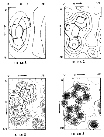
If some of the radiation scattered by an object under examination with a
microscope escapes rather than being recombined to form an image, the image so
formed will be an imperfect representation of the scattering object. Fine
detail will remain unresolved. Similarly, with X-rays, if the diffraction
pattern for the customary wavelengths is observed only out to a relatively small
scattering angle, the resolution of the corresponding image reconstructed will
be low. Furthermore, the resolution will be limited by the wavelength chosen
even if the entire pattern is observed. The `resolution' obtained is usually
expressed in terms of the interplanar spacings ![]() ,corresponding to the maximum observed
,corresponding to the maximum observed ![]() values. The effect of changing
resolution on the appearance of an electron density map is shown in Fig. 5.
Often, with macromolecules, the order does not persist from unit cell to unit
cell to high resolution. This lack of high resolution may also be observed when
data are collected for crystals near their melting points.
values. The effect of changing
resolution on the appearance of an electron density map is shown in Fig. 5.
Often, with macromolecules, the order does not persist from unit cell to unit
cell to high resolution. This lack of high resolution may also be observed when
data are collected for crystals near their melting points.
Copyright © 1984, 1997 International Union of Crystallography
IUCr Webmaster