![[IUCr Home Page]](/iucr-top/logos/iucrhome.gif)
![[Commission Home Page]](/iucr-top/logos/cteach.gif)
![]()
![]()
![]()
Next: 2. Some Textbooks
Up: 1. General Teaching
Previous: a. 10 minute talk
1st hour . Diffraction is discussed, as above. If white light is used to produce the diffraction pattern, some splitting of the spots into red and blue is seen, and this effect can be used to explain the wavelength effect on the diffraction angle. It is then explained how the diffraction pattern is measured (detectors, cameras, geometry, etc.) and the types of diffraction patterns obtained (usually precession or oscillation photographs or diffractometer measurements). This can also be done with a model, such as a collimator stuck into a styrofoam brick (X-ray tube), a goniometer head with a dummy crystal mounted on a wire, and precession photograph (preferably Polaroid, mounted on a wooden stand). The heights of each should be appropriately adjusted so that an imaginary X-ray beam passes through the collimator, hits the crystal, and then falls on the centre of the film (marked to indicate a beam catch for the direct beam). The physics of diffraction and the concepts of path differences and orders of diffraction are then explained. Make sure at the end of this that the audience knows what a diffraction data set is.
2nd hour . Then the audience is told how structures are derived from the diffraction data set. First, the reason why some `reflections' are intense is explained in terms of structure (for example, the DNA photograph and model could be used). Then the Patterson map is described in detail. The best simple description is given by Judson in The Eighth Day of Creation . Imagine a party and that at a given instant everyone's shoes were stuck to the floor. If each person then shook hands with every other person how would he turn, how far must he stretch and in total how may interactions would there be? This introduces the concept of vectors very simply. It is helpful then to analyse the Patterson map of a simple structure and one containing a heavy atom. Also it is shown how the orientations of groups of known structure may be deduced by comparing (and reorienting if necessary), a calculated and observed Patterson map.
Then the use of direct methods is described for the simple (centrosymmetric)
case of the 1 0 0 and the 2 0 0 reflection (both intense, hence 2 0 0 has a
phase of 0![]() ) (see Glusker and Trueblood, Crystal Structure
Analysis: A Primer ). This explanation can be expanded to more general
reflections if the audience is sufficiently interested. Then the use of
isomorphous replacement is described. This can be illustrated elegantly by
optical diffractions (with a laser light) of repeating patterns reduced to a
suitably small size (for example, Sung Hou Kim uses a drawing of a duck, and of
various eggs as the different heavy atoms). The intensity variation on
isomorphous replacement (an egg vs . no egg) can then be seen. (See
Atlas of Optical Transforms and
Pamphlet No. 1
which describe experiments on optical diffraction in detail.)
) (see Glusker and Trueblood, Crystal Structure
Analysis: A Primer ). This explanation can be expanded to more general
reflections if the audience is sufficiently interested. Then the use of
isomorphous replacement is described. This can be illustrated elegantly by
optical diffractions (with a laser light) of repeating patterns reduced to a
suitably small size (for example, Sung Hou Kim uses a drawing of a duck, and of
various eggs as the different heavy atoms). The intensity variation on
isomorphous replacement (an egg vs . no egg) can then be seen. (See
Atlas of Optical Transforms and
Pamphlet No. 1
which describe experiments on optical diffraction in detail.)
3rd hour . This time is most effectively spent by repeating the second hour's lesson. Usually most students have understood by the second time around.
4th hour . The methods of refinement are described, i.e. difference maps and particularly the method of least squares (using a simple linear equation to illustrate this method graphically). The estimation of precision (not to be confused with accuracy) of the result is then described.
The types of results and the information in crystal structure publications,
particularly protein structure papers, are then described. One useful way to
help the student understand descriptions of protein folding is to have him
thread beads of different colours (one colour for ![]() -helix, another for
pleated sheet, etc.) on a string and then fold the string of beads as described
in the article.
-helix, another for
pleated sheet, etc.) on a string and then fold the string of beads as described
in the article.
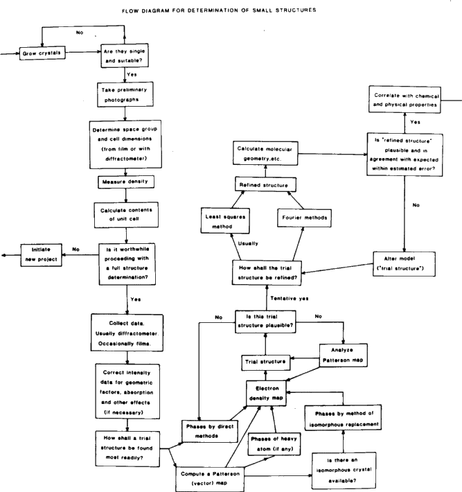
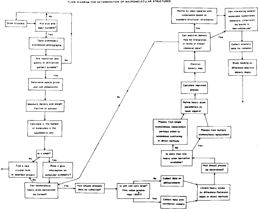
Some flow charts for structure determination are shown in Figs. 3 and 4.
Copyright © 1984, 1997 International Union of Crystallography
IUCr Webmaster