![[IUCr Home Page]](/iucr-top/logos/iucrhome.gif)
![[Commission Home Page]](/iucr-top/logos/cteach.gif)
![]()
![]()
![]()
Next: Lysozyme
Up: Elementary X-Ray Diffraction for Biologists
Previous: f. Comparisons of structures using the
Enzymes are polypeptides which use ordinary chemical mechanisms and specific binding interactions to speed up reactions. No mysterious forces need be invoked. Studies of the mechanisms by which enzymes catalyse reactions have been made by both biochemists and crystallographers, and it is when they work together that the most information is obtained. Once the three-dimensional structure has been determined by the crystallographer, further information may be obtained on the mechanism by NMR studies, chemical modification, and by X-ray crystallographic studies of enzyme-inhibitor complexes. The lock-and-key model of Emil Fischer (the enzyme is the lock and the substrate is the key), with some modification, is relevant to the interactions involved.
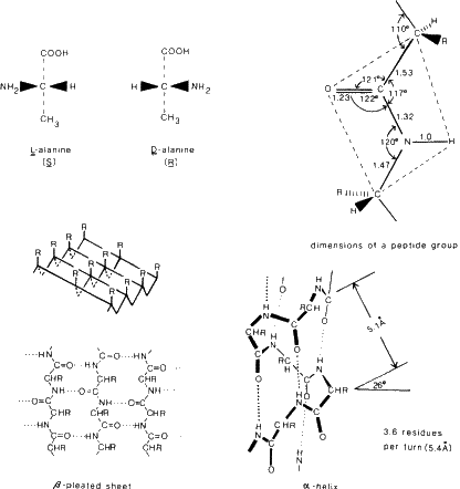
The building blocks are L-amino acids, NH+3--CHR--COO-,
where R is a substituent which defines the particular amino acid. There are 20
that are normally found in proteins and the structures of these have been
determined by X-ray and neutron diffraction methods. In proteins these amino
acids are joined by a peptide linkage C--NH--CO--C. Essentially this is a
planar grouping (as found in numerous peptides) and it is hinged to the next
peptide group at the carbon atom to give a flexible backbone composed of planar
peptide segments. Some dimensions are listed in Fig. 6. However two important
types of interactions can occur as the three-dimensional structure of a protein
is built up. One is hydrogen bond formation and the other is disulfide bond
formation. These interactions stabilize the molecular shape. In particular
hydrogen bond formation is responsible for the existence of ![]() -helices and
-helices and
![]() -pleated sheets, so common in enzymes.
-pleated sheets, so common in enzymes.
The main structural features of globular proteins may be generalized as follows (although exceptions are found):
General stereoviews, Dickerson, R. E. and Geis, I., The Structure and Action of Proteins . Menlo Park, California, Benjamin (1969).
When teaching about macromolecules be sure to look up recent issues of Nature, Proceedings of the National Academy of Sciences, the Journal of Molecular Biology and the Journal of Biological Chemistry for recent advances.
Copyright © 1984, 1997 International Union of Crystallography
IUCr Webmaster