Abstract Macromolecular crystallography (MX) continues to play a central role in atomic-resolution structural biology, even as the field undergoes rapid change driven by advances in complementary experimental and computational approaches. Techniques such as cryo-electron microscopy, neutron diffraction, spectroscopy, and deep-learning–based structure prediction and refinement are increasingly integrated with MX, enabling more comprehensive and quantitative descriptions of biomolecular structure and function.
This article synthesizes key insights, discussions, and representative case studies from the APS–NSLS-II–SLAC Joint Workshop held during the SLAC User Meeting on 26 September 2025, which brought together MX beamline scientists, method developers, computational researchers, and industrial users. The workshop highlighted progress in time-resolved and room-temperature crystallography, metallo-crystallography, membrane-protein structure determination, ultra-high-resolution studies, and AI-assisted analysis and refinement.
Discussions emphasized the growing importance of hybrid experimental strategies that combine X-ray diffraction with neutron and spectroscopic methods, together with AI-guided workflows for experiment design, data analysis, and interpretation. Looking forward, MX is expected to remain a foundational component of integrated, automated, and data-driven structural biology pipelines, contributing uniquely to precise ligand characterization, metal chemistry, hydrogen positioning, and dynamic studies. The perspectives summarized here outline key opportunities and challenges for MX facilities over the next decade as structural biology enters an increasingly multimodal and computationally integrated era.
- Introduction:
For more than seven decades, macromolecular crystallography (MX) has provided atomic-level insight into biological macromolecules and has remained a foundational technique in mechanistic biochemistry and structure-guided drug discovery, a legacy reflected in multiple Nobel Prizes (Jaskolski et al., 2014; Richardson & Richardson, 2014; Rathore et al., 2021). Despite the rapid growth of cryo-electron microscopy (cryo-EM), X-ray free-electron lasers (XFELs), and artificial-intelligence (AI)–based structure prediction, MX continues to provide uniquely precise experimental information, particularly for ligand binding, metal coordination, hydrogen placement, and ultra-high-resolution analysis.
Rather than being displaced by emerging approaches, MX is increasingly embedded within an integrative structural biology framework in which experimental and computational methods converge. Cryo-EM, neutron diffraction, spectroscopy, and predictive modeling now routinely complement crystallographic measurements, raising important questions about how MX facilities and user communities should evolve to maximize scientific impact. The key issue is therefore not whether MX will remain relevant, but how it will best synergize with complementary techniques to address increasingly complex biological problems.
The motivation for the present workshop arose from more than two decades of experience operating and developing MX facilities at U.S. synchrotrons, during which user priorities, experimental strategies, and publication patterns have changed substantially. Recent years have seen increasing demand for hybrid-method workflows, time-resolved (TR) and room-temperature (RT) experiments, automated data analysis, and closer integration with AI-driven modeling. These trends highlight the need for coordinated planning across facilities and for a reassessment of MX’s evolving role within the broader structural biology ecosystem.
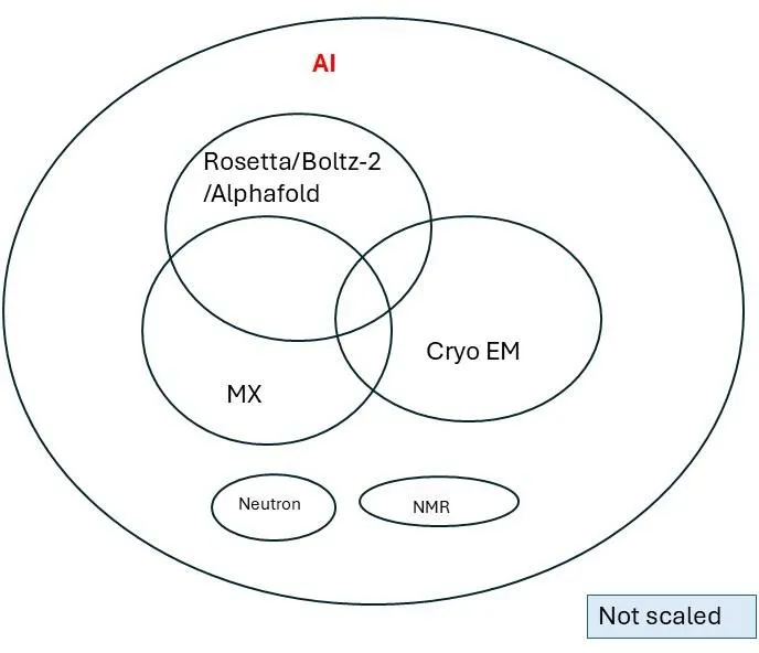
Figure 1 Core principle on which workshop was developed. MX: Macromolecular crystallography, AI: Artificial Intelligence.
The APS–NSLS-II–SLAC Joint Workshop (Fig. 1) was organized to address these challenges and opportunities by bringing together representatives from synchrotron MX, XFELs, cryo-EM, neutron scattering, spectroscopy, and computational structural biology. The Northeastern Collaborative Access Team (NECAT–APS), the Center for Biomolecular Structure (CBMS-NSLS-II), and the Linac Coherent Light Source (LCLS–SLAC) served as primary contributors, with additional participation from SSRL in various roles. It attracted ~100 registrations, including representatives from the DOE and NIH. The workshop aimed to assess the current state of MX, identify emerging synergies with complementary techniques, and outline strategic directions for facilities and user communities over the next decade. Details of speakers, chairs, and moderators are provided in Appendix 1.
This article summarizes the key discussions and outcomes of the workshop. Section 2 reviews facility capabilities and community perspectives presented during the meeting. Section 3 synthesizes major scientific and strategic insights, while Section 4 outlines recommendations for future development. The final sections discuss anticipated impacts and broader implications for the next generation of integrated structural biology.
- MX facilities:
Beamline management teams from facilities across the U.S.—from the East to the West Coast—presented spotlight overviews of the unique capabilities and future developments at their respective centers. The summary of U.S. macromolecular crystallography (MX) facilities highlights how synchrotron and XFEL centers (APS, NSLS-II, LCLS, MacCHESS, ALS, and SSRL) are collectively evolving toward an integrated, AI-driven, multi-technique ecosystem for structural biology. Historically, synchrotrons revolutionized the field by delivering atomic-resolution insights that enabled mechanistic discovery, supported structure-guided drug design, and generated the rich structural datasets that later fueled advances in AI and machine learning. Today, MX remains essential for high-resolution protein–ligand complexes and pharmaceutical applications, even as it increasingly complements cryo EM and predictive modeling rather than competing directly with them. Across U.S. facilities, capabilities continue to expand through microfocus MX, variable environmental (temperature, pressure and humidity) crystallography, fragment-based screening for drug discovery, and serial crystallography. These developments are tightly coupled with growing automation, AI-assisted data processing, and hybrid workflows that integrate multiple experimental and computational methods. Looking ahead, all major light sources emphasize cross-disciplinary synergy—positioning MX as a core component of broader structural and chemical biology programs. By integrating MX with cryo-EM, SAXS, spectroscopy, XFEL methods, and AI-driven tools, facilities aim to accelerate discovery across biology, chemistry, and materials science.
2.1 Summary of workshop and Community Perspectives
- Welcome
The workshop opened with remarks by Sébastien Boulet (LCLS) and Narayanasami Sukumar (NECAT-APS), who outlined expectations, anticipated outcomes, and strategies aimed at increasing the impact and visibility of macromolecular crystallography (MX). The meeting brought together MX beamline managers, computational scientists, method developers, and industrial researchers to assess the current landscape and define priorities for the future. The program was organized around four themed sessions that addressed strategic directions for U.S. MX facilities, advances in computation and the integration of artificial intelligence, the growing role of multimodal and non-X-ray techniques, and emerging frontiers in crystallography, including methodological innovations, scientific applications, and industry perspectives. The workshop concluded with a session focused on key scientific challenges, followed by a forward-looking round-table discussion.
- Session Summaries
2.1 Session 1: Strategic Directions for U.S. MX Facilities- Chair: Vivian Stojanoff (NSLS-II)
The opening scientific and spotlight presentations provided a global view of macromolecular crystallography (MX) capabilities and highlighted several critical future directions for U.S. facilities. These included advances in functional and dynamic studies; multi-crystal and multi-dataset strategies; time-resolved methodologies; RT crystallography; metal identification and metallo-crystallography; ultra-high-resolution studies; and expanded industrial applications. Additional emphasis was placed on the curation and effective use of large-scale datasets, enhanced training of early-career scientists, and the integration of synchrotron MX with cryo-EM for tissue-level and in vivo structural studies. Speakers also discussed the implementation of 4D/5D imaging, new opportunities enabled by the APS Upgrade, the need for greater standardization and dataset diversity, and the growing importance of automation, experimental flexibility, and cross-facility equipment sharing. Emerging complementarities between XFELs and synchrotron sources, particularly in serial crystallography approaches, were also highlighted.
A panel discussion moderated by Vivian Stojanoff (NSLS-II), James Baxter (LCLS), and Sébastien Boutet (LCLS) focused on strategies to expand the MX user base through improved training, enhanced university outreach, streamlined access mechanisms, and more strategic engagement with scientific communities. Panelists further emphasized the importance of developing improved facility-impact metrics, establishing coherent national data policies, and pursuing DOE-led initiatives for centralized metadata storage.
2.2 Session 2: Computational Advances and AI Integration- Chair: Clyde Smith (SSRL)
Talks in this session addressed rapid developments in AI-enabled structural biology, covering a range of topics that illustrated both recent progress and remaining challenges. Presentations discussed AI-driven enzyme design workflows, along with their current limitations in achieving high catalytic efficiency, and introduced Boltz-2, an open-source binding-affinity prediction tool, with comparisons to AlphaFold3. Speakers also highlighted ongoing challenges in crystallization prediction, particularly those arising from incomplete metadata and the lack of reported negative results. Advances in AI-guided refinement, troubleshooting, and quantum-refinement frameworks within the Phenix platform were presented, as were AI-supported approaches for the discovery and characterization of intrinsically disordered proteins, including the identification of new structural motifs in amyloid biology and microbial cellulosomes. Collectively, the session underscored the increasing integration of predictive modeling, crystallographic data, and automated pipelines in modern structural biology.
2. 3 Session 3: Multimodal and non-X-ray Techniques - Chair: Jennifer Wierman (MacCHESS)
This session focused on emerging and complementary structural biology tools, highlighting current and future developments in cryo-EM and cryo-electron tomography (cryo-ET) capabilities, along with national training efforts supporting their broader adoption.
Speakers compared the respective strengths of cryo-EM, cryo-ET, and microED for studies of large, intermediate, and small molecules, and discussed the unique contributions of neutron crystallography for probing hydrogen positions, protonation states, and catalytic mechanisms. The session also examined the role of solid-state NMR (ssNMR) in studies of membrane proteins, kinases, and ion channels, emphasizing its ability to reveal previously unseen conformational states.
A combined panel session spanning Sessions 2 and 3, moderated by Aina Cohen (SSRL) and James Holton (ALS), underscored recent advances in drug-design applications, the growing importance of multimethod integration, and the need for enhanced cross-disciplinary communication and training.
2.4 Session 4: Frontiers in Crystallography and Industry Perspectives- Chair: Sean McSweeney (NSLS-II)
Talks in this session addressed the integration of time-resolved crystallography with cryo-EM and AI/ML approaches, as well as the development of serial crystallography pipelines spanning timescales from femtoseconds to milliseconds. Speakers highlighted the complementary roles of XFELs in capturing dynamic processes, alongside MX, cryo-EM, and NMR in defining ground-state structural landscapes. Metallo-crystallography and ultra-high-resolution studies were also discussed, demonstrating how such approaches can reveal subtle differences in oxidation states.
Industry speakers emphasized the essential role of beamline scientists in maintaining productivity and efficiency, reaffirmed the continued value of X-ray crystallography for rapid drug–target characterization, and noted the expanding role of cryo-EM as throughput continues to improve. They further stressed the growing need for automation, machine-learning (ML) and deep-learning (DL) –based analytics, and high-throughput experimental pipelines to meet industrial demands.
2.5 Session 5: Scientific Challenges and Case Studies- Chair: Frank Murphy (NECAT-APS)
Talks in this session highlighted longstanding bottlenecks in membrane protein structural biology and presented new stabilization strategies that now enable near-atomic resolution structures of small and unstable membrane proteins, such as human vitamin K epoxide reductase. Speakers also compared the relative strengths of X-ray crystallography and cryo-EM for ribosomal studies, with prediction of continued dominance of cryo-EM in this area. In addition, the concept of statistical structural biology was discussed, integrating MX, cryo-EM, and AI approaches, including PanDDA-based methods for detecting low-occupancy ligands and the potential for developing high-throughput cryo-EM workflows.
The final roundtable, moderated by Narayanasami Sukumar (NECAT-APS), Vivian Stojanoff (NSLS-II), and Sébastien Boutet (LCLS), addressed the declining industrial emphasis on ribosome-targeting antibiotics, persistent challenges in modeling low-occupancy ligands, limitations of current ML/DL-based affinity prediction methods, and the growing need for MX facilities to clearly distinguish between high-throughput service pipelines and highly specialized experimental missions.
- Outcomes and Strategic Directions from the Workshop
Macromolecular crystallography (MX) continues to offer unique strengths, including sub-angstrom resolution, reliable hydrogen identification, detailed solvent mapping, and precise characterization of metal coordination chemistry. Persistent misconceptions that AI or cryo-EM can replace MX highlight the need for improved user education and outreach, while the long-term sustainability of MX will depend on deeper hybridization with complementary modalities, increased automation, and rigorous cross-method validation. MX and cryo-EM together provide hierarchical and complementary structural insights, with MX refining atomic-level details and cryo-EM capturing larger assemblies and broader conformational landscapes, whereas neutron diffraction and spectroscopic techniques such as XANES/EXAFS and micro-spectrophotometry supply critical protonation, oxidation-state, and chemical information not accessible through X-rays alone. Upgraded storage rings and AI-assisted pipelines are expected to further enhance productivity, throughput, and reproducibility across these methods.
Hybrid, AI-enabled, and time-resolved approaches are increasingly central to modern structural biology. Integrated crystallography–spectroscopy experiments continue to elucidate complex metalloprotein redox mechanisms, while advances in microfocus MX have enabled studies of challenging intrinsically disordered protein systems, including amyloid motifs, with cryo-EM and NMR providing validation of structural ensembles. Time-resolved XFEL MX, TR cryo-EM, and ML –based analyses now allow protein motions to be probed across timescales ranging from femtoseconds to milliseconds. In drug discovery, high-resolution MX is expected to remain essential for defining ligand geometry, hydrogen-bonding networks, and fragment screening, while cryo-EM and MicroED complement MX for large complexes, membrane proteins, and microcrystalline samples; automation is expected to increasingly shape workflow for target assessment, hit identification, and structure–activity relationship optimization.
Innovation in membrane-protein structural biology is being driven by terminus-restrained stabilization strategies that improve expression and crystallizability, alongside mass spectrometry and AlphaFold-guided construct design approaches that are enhancing success rates across GPCRs, ion channels, and transporters. In parallel, AI and ML tools are becoming integral to model building and refinement, with platforms such as Phenix incorporating AI assistants and quantum-refinement modules that improve both MX and cryo-EM structures, particularly at moderate resolution. Although predictive tools such as Boltz-2, AlphaFold and Rosetta generate powerful, testable hypotheses, they remain complementary to, rather than replacements for experimental validation. Meeting future scientific demands will require expanded capabilities in serial MX, RT data collection, spectroscopy, fragment screening, and variable-environment experiments, supported by nationally coordinated training programs to develop a technically confident and methodologically versatile workforce. Looking ahead, deeper integration of synchrotron, XFEL, cryo-EM, neutron scattering, and AI platforms is expected to shape the future of structural biology, necessitating unified interfaces, standardized metadata, and shared data formats to support reproducibility, accessibility, and efficient multimodal workflows.
- Recommendations for the Next Decade
The workshop clarified both the limitations and opportunities that are likely to shape macromolecular crystallography (MX) over the next five to ten years, with recommendations organized across several interconnected domains. In the area of data policies, access, and interoperability, participants emphasized the need for harmonized national data policies across DOE, NIH, and NSF to enable effective data sharing, support large language models for ML, and improve the overall efficiency of national user facilities. Sustained federal funding over long-time horizons was identified as essential for instrument innovation and the development of unique experimental components, particularly given that many enabling technologies are driven by private companies outside the United States. The expansion of the Universal Proposal System was recommended to better track scientific outcomes, patents, industrial engagement, and project movement across synchrotrons, XFELs, cryo-EM centers, and neutron facilities, alongside the harmonization of safety, shipping, and sample-handling procedures to provide a consistent and user-friendly experience.
With respect to facility coordination, scheduling, and strategic identity, the workshop highlighted the importance of stronger synchrony among synchrotrons, XFELs, and cryo-EM centers for sample delivery, data processing, and technology transfer. Participants encouraged exploration of synchrotron upgrades that would enable XFEL-like capabilities, particularly for dynamic and TR experiments, and stressed the need for MX beamlines to articulate clear strategic missions, whether focused on high-throughput pipelines or specialized, complex experiments—to reduce redundancy and enhance national complementarity. Maintaining coordinated national scheduling and operational redundancy was also seen as critical to ensuring reliable user access across major light sources.
Training, workforce development, and user support emerged as another central theme. Recommendations included enhanced user training that combines robust remote instruction with targeted in-person experiences for advanced, serial, TR, and multimodal experiments, as well as the adoption of multidisciplinary proposal review processes that incorporate expertise from cryo-EM, NMR, neutron scattering, and computational biology. In parallel, discussions on experimental methodology and technological development underscored that while most MX experiments do not require variable energy, energy tunability remains essential for metallo-enzymes and ultra-high-resolution studies. Participants advocated for broader adoption of serial crystallography to mitigate radiation damage, the provision of optional kappa goniometers to improve reciprocal-space coverage and dose efficiency, and the maintenance of flexible beam-size options to accommodate both microcrystals and larger samples. Strengthening pipelines for metallo-crystallography and low-occupancy ligand discovery, expanding multimodal data-deposition frameworks that integrate X-ray, cryo-EM, neutron, and spectroscopic data, and increasing engagement with catalysis, materials science, and molecular engineering communities were also identified as priorities.
Finally, the workshop highlighted the transformative role of AI, automation, and predictive structural biology. Participants recommended deeper integration of AI into facility operations for experiment design, automated data acquisition, real-time diagnostics, and preliminary interpretation, as well as the development of AI-driven crystallization-prediction models trained on literature, PDB metadata, and experimental outcomes to reduce longstanding bottlenecks. The integration of structural databases with biochemical and medicinal chemistry data was seen as a pathway toward AI-enhanced structure–activity relationship pipelines, while AI-driven beam optimization was identified to improve stability, calibration, and ease of use. Throughout these discussions, the importance of human–AI collaboration was emphasized, with training programs needed to ensure that early-career researchers can critically interpret AI-assisted results and understand their limitations. Lowering barriers between experimental modalities through automation and AI was also viewed as essential for enabling seamless transitions among MX, cryo-EM, MicroED, and spectroscopic approaches and fostering broader cross-disciplinary adoption.
- Predictive Next-Generation Structural Biology at MX Facilities
This session outlined forward-looking and actionable trajectories for macromolecular crystallography over the next five to ten years, emphasizing the importance of focused and strategic planning. A central theme was the emergence of AI-driven, fully automated MX pipelines, in which end-to-end “cradle-to-PDB” workflows—encompassing crystal centering, data collection, processing, refinement, and validation—will increasingly incorporate large language model–guided decision tools. AlphaFold- and LLM-enabled molecular replacement, quantum-assisted refinement, and AI-based quality assessment are expected to shift human effort away from routine analysis toward deeper biological interpretation. In parallel, integrated spectroscopy and diffraction will become standard practice, with techniques such as XANES/EXAFS, UV–Vis, and Raman spectroscopy embedded directly into beamlines and automatically triggered to probe oxidation states, photochemistry, and chromophore dynamics during diffraction experiments.
Technological advances will further be driven by the adoption of dual-detector architectures, combining GaAs detectors operating in the 8–20 keV range with CdTe detectors above 20 keV to improve data quality, reduce radiation damage, and expand accessible experimental parameter space. Serial and TR crystallography are expected to become mainstream, as fourth-generation sources routinely support MHz-rate serial data collection and dynamic experiments once limited to XFELs. TR studies will span femtosecond to millisecond timescales at XFELs and microsecond to second regimes at synchrotrons, enabling the generation of detailed molecular “movies.” Routine RT data collection and variable-environment experiments will be enabled by faster shutterless acquisition and serial strategies, while standardized microfluidic chips will allow controlled variation of pressure, humidity, temperature, ligands, electric fields, and photoactivation during experiments.
Radiation-damage mitigation will remain a priority, with expanded use of high-energy crystallography in the 18–35 keV range coupled to real-time dose monitoring, alongside widespread adoption of multi-crystal and serial “diffraction-before-damage” approaches. XFEL developments, including LCLS-HE upgrades, will enable MHz-rate serial femtosecond crystallography at 25–30 keV, while harmonized sample stages will simplify planning and execution of coordinated multi-facility experiments. Advanced multi-color, two-pulse pump–probe schemes are expected to provide new insights into ultrafast charge migration and electron dynamics.
The session also emphasized expansion beyond classical structural biology, with MX and serial methods increasingly applied to materials science, catalysis, polymer chemistry, and metal–organic framework research. Integrated PDB- and CSD-style archives are envisioned to unify macromolecular and small-molecule crystallography with shared multimodal metadata. Workforce development will evolve in parallel, with hybrid fellowships spanning MX, cryo-EM, spectroscopy, and computation becoming standard training pathways, and beamline graphical interfaces incorporating AI-guided tutorials to lower barriers for users from diverse scientific backgrounds.
Quantitative milestones were proposed to benchmark progress, including expectations that more than half of MX-derived PDB entries will incorporate AI-assisted refinement, that over 30% of MX beamtime will be devoted to serial and time-resolved methods, and that median crystal sizes will fall below 10 μm as microcrystal-compatible approaches mature. Rapid ligand and fragment structure determination within hours is anticipated to become routine. Complementary techniques such as cryo-EM, cryo-ET, and MicroED will continue advancing toward higher resolution and faster temporal domains, supported by next-generation foundation models, including future iterations of Rosetta, Boltz, and AlphaFold—that generate dynamic structural ensembles with ligand-confidence metrics. Shared data formats, sample supports, and AI co-pilots will streamline multimodal experiments, accelerating a shift from technique-centered projects to integrated, contrast-combined campaigns.
Finally, the session underscored the need for a comprehensive impact matrix that extends beyond traditional metrics such as publications, PDB depositions, and user numbers to include patents, translational outcomes, and pre-competitive industrial collaborations. Because modern structural biology is inherently integrated and multimodal, such tracking should capture how projects progress across synchrotrons, XFELs, cryo-EM, and neutron facilities, while also evaluating interoperability through harmonized sample handling, metadata standards, proposal systems, and coordinated training. This level of coordination was identified as essential for enabling seamless multimodal research and supporting a truly user-centered national infrastructure.
- Conclusions
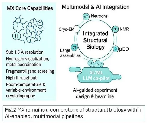
Figure 2 MX remains a cornerstone of structural biology within AI-enabled, multimodal pipelines
Macromolecular crystallography remains a cornerstone of structural biology, defined by its unmatched precision in atomic resolution, hydrogen visualization, solvent structure, and metal coordination. The workshop affirmed that MX is now embedded within a broader, highly interconnected ecosystem that includes cryo-EM, MicroED, neutron scattering, spectroscopy, XFEL science, and rapidly advancing AI-driven computation. In this evolving landscape, MX serves as a central pillar within coordinated, multimodal structural investigations—providing types of data and experimental capabilities that no other method can match in accuracy, throughput, or scalability (Fig. 2).
Participants identified the coming decade as a period of rapid transformation shaped by AI-enabled automation, methodological complementarity, and system-level coordination across U.S. national laboratories. Achieving this vision will require unified national data policies, interoperable standards for multimodal experimentation, and coordinated scheduling and access across facilities. As AI increasingly guides experiment design, beamline optimization, data processing, refinement, and interpretation, scientists will be able to devote their expertise to mechanistic insight and hypothesis-driven discovery.
The workshop also articulated a clear scientific identity for MX centered on areas where it provides unique and irreplaceable value: RT and variable-environment crystallography; ultra–high-resolution structural analysis; precise hydrogen placement and protonation-state assignment; detailed solvent and ligand mapping; high-precision metalloprotein characterization; and fragment/ ligand screening at throughput unmatched by any other technique. TR MX—from femtoseconds at XFELs to seconds at synchrotrons—remains unique in its ability to visualize conformational dynamics across an exceptionally broad temporal range. Emerging advances in automation, high-energy beamline operation, dual-detector geometries, and integrated spectroscopy will further expand these strengths and reinforce MX’s singular contributions.
Equally important, the workshop emphasized that MX’s unparalleled efficiency and scale will be essential in the data-driven era of structural AI. As computational models increasingly require curated, high-quality, and chemically accurate training data, MX will continue to provide the large, diverse, and experimentally validated structural datasets needed to advance predictive biology.
A strong consensus emphasized the essential role of workforce development. As experimental platforms become increasingly automated and multimodal, researchers, especially early-career scientists, must be trained to interpret data judiciously, understand method-specific limitations, and use AI tools in responsible and scientifically rigorous ways. Hybrid training models that span MX, cryo-EM, and computation will be critical to building a workforce capable of leading next-generation structural science.
Finally, sustaining U.S. leadership in structural biology will require strategic investment, facility-to-facility collaboration, and broad community engagement. By integrating AI, advancing multimodal pipelines, strengthening interoperability across facilities, and focusing on the areas where MX provides unique scientific leverage, the field is exceptionally well positioned for the future. In an era of rapidly expanding methodologies, MX will remain a foundational, quantitative, and experimentally rigorous technique—anchoring the next generation of predictive, dynamic, and truly multimodal structural biology, while continuing to deliver insights at a scale and precision unmatched by any other approach.
Acknowledgements:
The authors acknowledge valuable contribution of speakers, chairs and moderators of the 2025 APS–NSLS-II–SLAC Joint Workshop (Listed in Appendix 1) . Funding support from the SLAC (LCLS/SSRL user committee) is gratefully acknowledged. The organizers, Narayanasami Sukumar is part of NECAT (APS-Cornell University), which is supported by NIH-P30 GM124165, Vivian Stojanoff, is part of the Center for Biomolecular Structure, NSLS-II, and is supported by NIH-P30-GM133893 and BER FMP #BO070, while James Baxter and Sébastien Boutet are part of LCLS. The Linac Coherent Light Source (LCLS), SLAC National Accelerator Laboratory, is supported by the U.S. Department of Energy, Office of Science, Office of Basic Energy Sciences under Contract No. DE-AC02-76SF00515. This work was supported by NIH grant P41GM139687.
Appendix 1
| Narayanasami Sukumar (NECAT-APS) | Organizer | Speaker | Moderator |
| Vivian Stojanoff (CBMS-NSLS-II) | Organizer | Moderator | |
| Sébastien Boutet (LCLS) | Organizer | Moderator | |
| James Baxter (LCLS) | Co-organizer | Speaker | Moderator |
| James Holton (ALS) | Speaker | Moderator | |
| Sean McSweeney (NSLS-II) | Speaker | Chair | |
| Jennifer Wierman (MacCHESS) | Speaker | Chair | |
| Frank Murphy (NECAT-APS) | Speaker | Chair | |
| Greg Hura (ALS) | Speaker | ||
| Aina Cohen (SSRL) | Speaker | Moderator | |
| Leland Gee (LCLS) | Speaker | ||
| Sam Pellock (University of Washington) | Speaker | ||
| Jeremy Wohlwend (MIT) | Speaker | ||
| Sarah Bowman (HWI-Buffalo) | Speaker | ||
| Pavel Afonine (LBNL) | Speaker | ||
| Mike Sawaya (UCLA) | Speaker | ||
| Lydia-Marie Joubert (SSRL) | Speaker | ||
| Pete Dahlberg (Stanford,SSRL) | Speaker | ||
| Flora Meilleur (ORNL,North Carolina) | Speaker | ||
| Ann McDermott (Columbia) | Speaker | ||
| Marius Schmidt (UWM) | Speaker | ||
| Jim Kiefer (Genentech) | Speaker | ||
| Sandra Gabelli (Merck) | Speaker | ||
| Weikai Li (WashU, STL) | Speaker | ||
| Yury Polikanov (UIC) | Speaker | ||
| James Fraser (UCSF) | Speaker |
Author Information
Narayanasami Sukumar1*, Vivian Stojanoff2*, James Baxter3, Sébastien Boutet3*
1NE-CAT, Department of Chemistry and Chemical Biology, Cornell University, Building 436E, Argonne National Laboratory, Argonne, IL 60439, USA
2Center of Biomolecular Structure, NSLS-II, Building 745, Brookhaven National Laboratory, Upton, NY 11973, USA
3LCLS, SLAC National Accelerator Laboratory, Menlo Park, CA 94025, USA
*Correspondence: [email protected], [email protected], [email protected]
References
Jaskolski, M., Dauter, Z. & Wlodawer, A. (2014). “A brief history of macromolecular crystallography, illustrated by a family tree and its Nobel fruits.” The FEBS Journal 281, 3985–4009. https://doi.org/10.1111/febs.12796
Rathore, I., Mishra, V. & Bhaumik, P. (2021). “Advancements in macromolecular crystallography: from past to present.” Emerg. Top. Life Sci. 5, 127–149. https://doi.org/10.1042/ETLS20200316
Richardson, J. S. & Richardson, D. C. (2014). “Biophysical Highlights from 54 Years of Macromolecular Crystallography.” Biophysical Journal 106, 510–525. https://doi.org/10.1016/j.bpj.2014.01.001