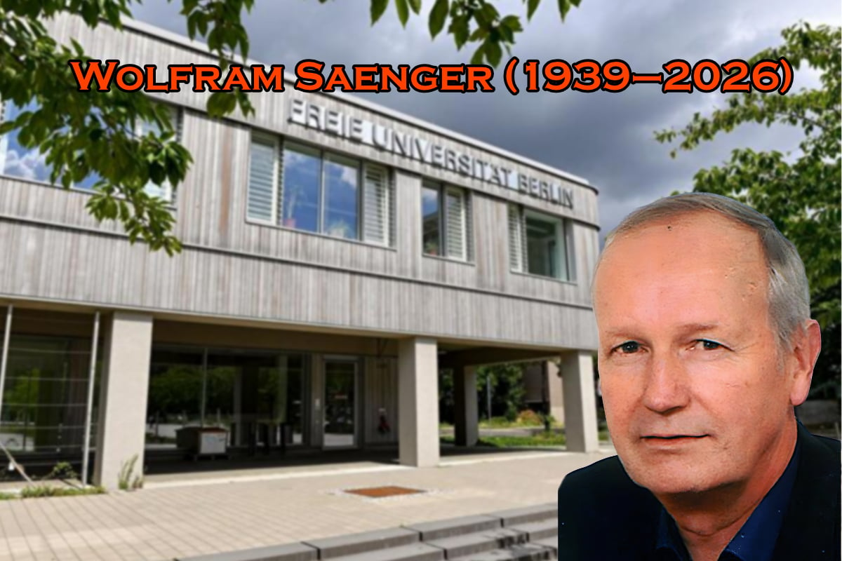
The international crystallographic community mourns the passing of Wolfram Saenger, a pioneering structural biologist whose work profoundly shaped our understanding of
nucleic acids, carbohydrates, protein–nucleic acid interactions and the structural basis of eukaryotic photosynthesis. Over a career spanning more than five decades, Saenger combined rigorous crystallographic analysis with theoretical insight and an unusually broad scientific vision, leaving a lasting legacy in structural biology.
Wolfram Saenger was born on 23 April 1939 in Frankfurt-Höchst, Germany. His father was an industrial chemist, and the family relocated several times during his childhood. Saenger spent his school years in Grenzach on the Rhine opposite Basel. From 1958 to 1964 he studied chemistry at the Technische Hochschule Darmstadt and at the University of Heidelberg. In 1964 he obtained his Diplom at TH Darmstadt. One year later, he completed his doctoral degree (Dr.-Ing.) under the supervision of Friedrich Cramer, studying rapid kinetics of cyclodextrin inclusion complexes in the laboratory of Manfred Eigen in Göttingen (Cramer et al., 1967).
From 1965 to 1967 Saenger pursued postdoctoral research at Harvard University, working with Robert B. Woodward and Jack Z. Gougoutas, where he learned X-ray crystallographic methods for small organic molecules (Simon et al., 1967). During this period he also collaborated with Elias J. Corey, including work on the symmetric synthesis of α-amino acids that appeared in 1970 (Corey et al., 1970).
Saenger returned to Germany in 1967 to head the X-ray crystallography group in the Department of Chemistry at the Max Planck Institute for Experimental Medicine in Göttingen, directed by Friedrich Cramer. He completed his habilitation at the University of Göttingen in 1971. After receiving several offers in the late 1970s, he accepted a professorship in 1981 at the Institute of Crystallography of Freie University of Berlin, where he built an internationally renowned centre for structural biology.
Saenger's early scientific interests focused on host–guest chemistry, particularly the structures and inclusion complexes of cyclodextrins and polyethers (Manor & Saenger, 1972; Hingerty & Saenger, 1975; Noltemeyer & Saenger, 1976; Gessler et al., 1994). The subtle and highly specific interactions between host and guest molecules provided model systems for understanding molecular recognition in biology. These studies naturally led him to the central role of hydrogen bonding in biomolecular structure. In cyclodextrin structures, he recognized the presence of three-dimensional hydrogen-bonding networks with circular arrangements of four or more water molecules (Saenger, 1979) and the cooperative flip of H atoms in equilibrium between two states: O—H⋯O ⇌ O⋯H—O (Saenger et al., 1982). Together with William N. Hunter and Olga Kennard, he recognized the importance of hydrogen bonding and hydration on the conformation of DNA (Saenger et al., 1986). His work in this area culminated in the influential monograph Hydrogen Bonding in Biological Structures, written together with George A. Jeffrey, which remains a foundational reference (Jeffrey & Saenger, 1991).
Cyclodextrins and other oligosaccharide and polysaccharides remained a recurring theme in Saenger's research (Hinrichs et al., 1987). Among his notable achievements was the structural characterization of a cyclic oligoglucose consisting of 26 monomer units (Gessler et al., 1999). Yet from early in his career he was equally fascinated by nucleic acids and their building blocks (Suck et al., 1972; Saenger, 1973; Saenger & Suck, 1973; Saenger et al., 1977). As early as 1968 he reported single crystals of phenylalanine-specific transfer RNA (Cramer et al., 1968), placing him and Friedrich Cramer in the international race to determine the first tRNA structure. Saenger's work on modified nucleic acid bases, abundant in tRNA, included landmark studies such as the structure of the base pair between 1-methyl-4-thiouracil and 9-methyladenine published in Nature in 1970 (Saenger & Suck, 1970).
Beyond experimental crystallography, Saenger increasingly integrated theoretical approaches into his studies of nucleosides and nucleotides, collaborating with leading theoreticians including Bogdan Lesyng and Camillo A. Tosi (Banerjee et al., 1978). His deep and comprehensive knowledge of nucleic acid structure was synthesized in his widely acclaimed book Principles of Nucleic Acid Structure (Saenger, 1983), which became a standard reference for students and researchers alike.
During his later years in Göttingen and especially after moving to Berlin, Saenger expanded his work decisively into protein crystallography. The first protein structures determined in his laboratory was that of α-cobratoxin (Walkinshaw et al., 1980), whose interaction with the nicotinic acetylcholine receptor provided important insights into receptor binding. Soon afterwards, his group solved the structure of the enzyme ribonuclease T1 (Heinemann & Saenger, 1982), initiating a long-standing programme combining crystallography with spectroscopy, mutagenesis and computational modelling to explore enzyme function and stability.
In Berlin, Saenger's laboratory investigated a wide array of biologically important macromolecules. Central among these were proteins interacting with carbohydrates (Holzenburg et al., 1987) and protein–nucleic acid complexes, including the factor for inversion stimulation (FIS; Kostrewa et al., 1991), the tetracycline repressor (TetR; Hinrichs et al., 1994) and DNA methyltransferases involved in DNA repair and restriction–modification systems (Labahn et al., 1994; Schluckebier et al., 1997). These studies significantly advanced the structural understanding of gene regulation and DNA recognition.
A particularly ambitious line of research addressed large membrane-protein complexes. Saenger and his collaborators determined important structures of cyanobacterial photosystems I (Krauss et al., 1993, 1996; Jordan et al., 2001) and II (Zouni et al., 2001; Loll et al., 2005; Yano et al., 2006; Guskov et al., 2009), among the most challenging targets in structural biology at the time. Later projects extended to proteins of the human immune system, notably major histocompatibility complex (MHC) molecules that present antigen-derived peptides to T cells and thereby initiate cellular immune responses (Menssen et al., 1999; Uchanska-Ziegler et al., 2012).
Throughout his career Saenger was known for tackling problems at the limits of technical feasibility. Membrane proteins and large multi-subunit complexes, often considered among the most difficult targets in crystallography, featured prominently in his research. His ability to sustain such ambitious projects over many years reflected both scientific determination and a remarkable capacity to integrate diverse experimental and theoretical approaches.
Saenger's scientific productivity was extraordinary. He authored more than 600 scientific publications, many in leading journals, as well as several influential books and review articles. His work was widely recognized through numerous honours. He was elected a member of the European Molecular Biology Organization (EMBO) in 1984, received the Gottfried Wilhelm Leibniz Prize in 1988 and the Alexander von Humboldt Research Award in 1989, and became a member of the Berlin-Brandenburg Academy of Sciences and Humanities in 1994. In 2004 he received the Carl-Hermann Medal of the German Association of Crystallography for his life's work.
Saenger was head of the West German Arbeitsgemeinschaft Kristallographie and played an important role in the unification of crystallographers and the establishment of the German Crystallographic Society in 1991 after German reunification. He served as co-editor of Acta Crystallographica Section D and as chairperson of the Commission on Biological Macromolecules of the IUCr from 1996 to 1999. An important topic at that time was the deposition and release of the coordinates and diffraction data of macromolecular structures (Baker & Saenger, 1999). Wolfram Saenger played a leading role in establishing the Protein Structure Factory, a large-scale structural genomics initiative based in Berlin (Heinemann et al., 2000). The project led to the development of key research infrastructure, including synchrotron beamlines at BESSY II (Helmholtz Zentrum Berlin).
Equally important was Saenger's role as a mentor. Generations of students and postdoctoral researchers benefited from the intellectual freedom and generous support he offered. Many of his former group members have gone on to leading positions in academia and industry, forming what colleagues often described as a distinctive `Saenger school' of structural biology.
Wolfram Saenger believed that scientific research should be pursued with intellectual honesty, curiosity and uncompromising standards of quality. His work exemplified a rare breadth of interests, spanning small molecules, nucleic acids, proteins and large macromolecular assemblies, combined with a deep commitment to fundamental understanding. His contributions have left an enduring imprint on crystallography and structural biology, and his influence will continue to be felt through the many scientists that he trained and inspired.
This article was originally published in Acta Cryst. (2026). D82, 571–573.
References
Baker, E. N. & Saenger, W. (1999). Acta Cryst. D55, 2–3.
Banerjee, A., Saenger, W., Lesyng, B., Kazimierczuk, Z. & Shugar, D. (1978). Acta Cryst. B34, 2472–2477.
Corey, E. J., Sachdev, H. S., Gougoutas, J. Z. & Saenger, W. (1970). J. Am. Chem. Soc. 92, 2488–2501.
Cramer, F., Haar, F., v, , d, , Saenger, W. & Schlimme, E. (1968). Angew. Chem. Int. Ed. Engl. 7, 895.
Cramer, F., Saenger, W. & Spatz, H.-C. (1967). J. Am. Chem. Soc. 89, 14–20.
Geßler, K., Krauß, N., Steiner, T., Betzel, C., Sandmann, C. & Saenger, W. (1994). Science 266, 1027–1029.
Gessler, K., Usón, I., Takaha, T., Krauss, N., Smith, S. M., Okada, S., Sheldrick, G. M. & Saenger, W. (1999). Proc. Natl Acad. Sci. USA 96, 4246–4251.
Guskov, A., Kern, J., Gabdulkhakov, A., Broser, M., Zouni, A. & Saenger, W. (2009). Nat. Struct. Mol. Biol. 16, 334–342.
Heinemann, U., Frevert, J., Hofmann, K., Illing, G., Maurer, C., Oschkinat, H. & Saenger, W. (2000). Prog. Biophys. Mol. Biol. 73, 347–362.
Heinemann, U. & Saenger, W. (1982). Nature 299, 27–31.
Hingerty, B. & Saenger, W. (1975). Nature 255, 396–397.
Hinrichs, W., Büttner, G., Steifa, M., Betzel, C., Zabel, V., Pfannemüller, B. & Saenger, W. (1987). Science 238, 205–208.
Hinrichs, W., Kisker, C., Düvel, M., Müller, A., Tovar, K., Hillen, W. & Saenger, W. (1994). Science 264, 418–420.
Holzenburg, A., Mayer, F., Harauz, G., van Heel, M., Tokuoka, R., Ishida, T., Harata, K., Pal, G. P. & Saenger, W. (1987). Nature 325, 730–732.
Jeffrey, G. A. & Saenger, W. (1991). Hydrogen Bonding in Biological Structures. Berlin, Heidelberg: Springer. GoogleScholar
Jordan, P., Fromme, P., Witt, H. T., Klukas, O., Saenger, W. & Krauß, N. (2001). Nature 411, 909–917.
Kostrewa, D., Granzin, J., Koch, C., Choe, H. W., Raghunathan, S., Wolf, W., Labahn, J., Kahmann, R. & Saenger, W. (1991). Nature 349, 178–180.
Krauß, N., Schubert, W. D., Klukas, O., Fromme, P., Witt, H. T. & Saenger, W. (1996). Nat. Struct. Mol. Biol. 3, 965–973.
Krauss, N., Hinrichs, W., Witt, I., Fromme, P., Pritzkow, W., Dauter, Z., Betzel, C., Wilson, K. S., Witt, H. T. & Saenger, W. (1993). Nature 361, 326–331.
Labahn, J., Granzin, J., Schluckebier, G., Robinson, D. P., Jack, W. E., Schildkraut, I. & Saenger, W. (1994). Proc. Natl Acad. Sci. USA 91, 10957–10961.
Loll, B., Kern, J., Saenger, W., Zouni, A. & Biesiadka, J. (2005). Nature 438, 1040–1044.
Manor, P. C. & Saenger, W. (1972). Nature 237, 392–393.
Menssen, R., Orth, P., Ziegler, A. & Saenger, W. (1999). J. Mol. Biol. 285, 645–653.
Noltemeyer, M. & Saenger, W. (1976). Nature 259, 629–632.
Saenger, W. (1973). Angew. Chem. Int. Ed. Engl. 12, 591–601.
Saenger, W. (1979). Nature 279, 343–344.
Saenger, W. (1983). Principles of Nucleic Acid Structure. New York: Springer-Verlag. GoogleScholar
Saenger, W., Betzel, C., Hingerty, B. & Brown, G. M. (1982). Nature 296, 581–583.
Saenger, W., Hunter, W. N. & Kennard, O. (1986). Nature 324, 385–388.
Saenger, W., Reddy, B. S., Mühlegger, K. & Weimann, G. (1977). Nature 267, 225–229.
Saenger, W. & Suck, D. (1970). Nature 227, 1046–1047.
Saenger, W. & Suck, D. (1973). Nature 242, 610–612.
Schluckebier, G., Kozak, M., Bleimling, N., Weinhold, E. & Saenger, W. (1997). J. Mol. Biol. 265, 56–67.
Simon, M. S., Rogers, J. B., Saenger, W. & Gougoutas, J. Z. (1967). J. Am. Chem. Soc. 89, 5838–5844.
Suck, D., Saenger, W. & VorbrÜggen, H. (1972). Nature 235, 333–334.
Uchanska-Ziegler, B., Loll, B., Fabian, H., Hee, C. S., Saenger, W. & Ziegler, A. (2012). Eur. J. Cell Biol. 91, 274–286.
Walkinshaw, M. D., Saenger, W. & Maelicke, A. (1980). Proc. Natl Acad. Sci. USA 77, 2400–2404.
Yano, J., Kern, J., Sauer, K., Latimer, M. J., Pushkar, Y., Biesiadka, J., Loll, B., Saenger, W., Messinger, J., Zouni, A. & Yachandra, V. K. (2006). Science 314, 821–825.
Zouni, A., Witt, H. T., Kern, J., Fromme, P., Krauss, N., Saenger, W. & Orth, P. (2001). Nature 409, 739–743.

![[Cover v25n2]](https://www.iucr.org/__data/assets/image/0018/135180/cover25n2.jpg)

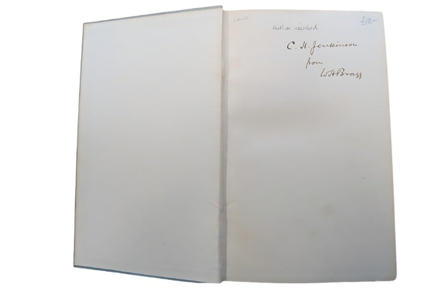 As some of you may be aware, I am always interested in the history of our subject. Recently, a colleague found in a second-hand bookshop in Oxford the book entitled The Universe of Light (1933) by Sir William Henry Bragg (WHB). WHB evidently gave this book to C.H. Jenkinson, who was WHB’s personal workshop technician, responsible for building the first ionisation spectrometer circa 1912, the forerunner of the modern X-ray diffractometer. The book was originally priced at four shillings and sixpence (about 23 UK new pence at the time).