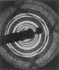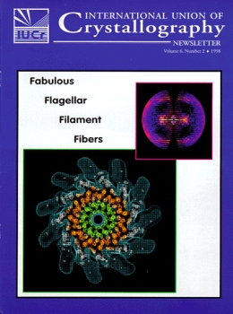
The X-ray fiber diffraction pattern from L and R-type flagellar filaments and the refined electron density map of the R-type filament (an end on view of a 50 Å thick cross section) are esthetically peasing and information rich. Bacteria swim by rotating flagellar filaments. During the straight swimming phase, the flagellar filaments form a bundle behind the cell body, where the filaments are all in a left-handed supercoiled form, each acting as a propellar driven by a rotary motor at the base of the filament. The flagellar filament is a tubular structure composed of 11 protofilament strands, which are axially aligned arrays of subunits. Bacterial motility involves switching between the left and right supercoiled states of the flagellar filament. The 9 Å resolution electron density map of the R-type filament, refined from the X-ray data, reveals details of the supercoil important to bacterial mobility.
Reference: I. Yamashita, K.Hasegawa, H. Suzuki, F. Vonderviszr, Y. Mimori-Kiyosue and K. Namba. Nature Structural Biology, Vol. 5, No. 2, 1998, pp 125-129.