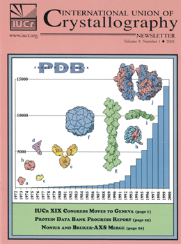
The number of structures in the PDB per year, as well as examples of structures in the PDB, are shown. In the 1970s, the first structures available to the scientific community included proteins such as myoglobin (a), hemoglobin (b) and lysozyme (c), and other molecules such as transfer RNA (d). In the 1980s, advances in experimental data collection allowed much larger structures to be solved, including antibodies (e) and entire viruses (f). In 2001, all aspects of structural science have advanced so that very complex structures are now accessible to study, including actin (g), the nucleosome (h), myosin (i), and even ribosomal subunits (j). Structures pictured here were taken from PDB entries 1mbn, 2dhb, 2lyz, 4tna + 6tna, 1fc1+1mcp, 2stv, 1atn, 1aoi, 1dfk, and 1ffk+1fka+1fjf, respectively. Images were created by David Goodsell of the Scripps Research Inst., who creates the PDB’s Molecule of the Month series.
- 1mbn: Watson, H. C.: The Stereochemistry of the Protein Myoglobin Prog.Stereochem. 4 pp. 299 (1969)
- 2dhb: Bolton, W., Perutz, M.F.: Three dimensional fourier synthesis of horse deoxyhaemoglobin at 2.8 Angstrom units resolution. Nature 228 pp. 551 (1970)
- 2lyz: Blake, C.C.F., Johnson, L.N., Mair, G.A., North, A.C.T., Phillips, D.C., Sarma, V.R.: Crystallographic Studies Of The Activity Of Hen Egg-White Lysozyme, Proc. R. Soc. London, Ser.B 167 pp. 378 (1967)
- 4tna: Hingerty, B., Brown, R.S., Jack, A.: Further refinement of the structure of yeast tRNAPhe. J Mol Biol 124 pp. 523 (1978)
- 6tna: Sussman, J.L., Holbrook, S.R., Warrant, R.W., Church, G.M., Kim, S.-H.: Crystal Structure of Yeast Phenylalanine T-RNA. I.Crystallographic Refinement J Mol Biol 123 pp. 607 (1978)
- 1fc1: Deisenhofer, J.: Crystallographic refinement and atomic models of a human Fc fragment and its complex with fragment B of protein A from Staphylococcus aureus at 2.9- and 2.8-A resolution. Biochemistry 20 pp. 2361 (1981)
- 1mcp: Satow, Y., Cohen, G.H., Padlan, E.A., Davies, D.R.: Phosphocholine binding immunoglobulin Fab McPC603. An X-ray diffraction study at 2.7 Å. J Mol Biol 190 pp. 593 (1986)
- 2stv: Jones, T.A., Liljas, L.: Structure of satellite tobacco necrosis virus after crystallographic refinement at 2.5 A resolution. J Mol Biol 177 pp. 735 (1984)
- 1atn: Kabsch, W., Mannherz, H.G., Suck, D., Pai, E.F., Holmes, K.C.: Atomic structure of the actin:DNase I complex. Nature 347 pp. 37 (1990)
- 1aoi: Luger, K., Mader, A.W., Richmond, R.K., Sargent, D.F., Richmond, T.J.: Crystal structure of the nucleosome core particle at 2.8 Å resolution. Nature 389 pp. 251 (1997)
- 1dfk: Houdusse, A., Szent-Gyorgyi, A.G., Cohen, C.: Three Conformational States of Scallop S1 Proc. Nat. Acad. Sci. USA 97 pp. 11238 (2000)
- 1ffk: Ban, N., Nissen, P., Hansen, J., Moore, P.B., Steitz, T.A.: The Complete Atomic Structure of the Large Ribosomal Subunit at 2.4 Å Resolution Science 289 pp. 905 (2000)
- 1fka: Schluenzen, F., Tocilj, A., Zarivach, R., Harms, J., Gluehmann, M., Janell, D., Bashan, A., Bartels, H., Agmon, I., Franceschi, F., Yonath, A.: Structure of Functionally Activated Small Ribosomal Subunit at 3.3 Å Resolution Cell 102 pp. 615 (2000)
- 1fjf: Wimberly, B.T., Brodersen, D.E., Clemons Jr., W.M., Morgan-Warren, R., Carter, A.P., Vonrhein, C., Hartsch, T., Ramakrishnan, V.: Structure of the 30S Ribosomal Subunit Nature 407 pp. 327 (2000)


