The challenges and potential for sub-atomic resolution X-ray crystallography and neutron crystallography
 The International Year of Crystallography saw the number of macromolecular structures deposited in the Protein Data Bank cross the 100,000 mark, with more than 90,000 of these provided by X-ray crystallography. The number of X-ray structures determined to sub-atomic resolution has passed 600 and this is likely to continue to grow rapidly with Diffraction Limited Synchrotron Radiation sources (DLSR) such as MAX IV (Sweden) and SIRIUS (Brazil) under construction. A dozen X-ray structures have been deposited to ultra-high resolution for which precise electron density can be exploited to obtain charge density and provide information on the bonding character of catalytic or electron transfer sites.
Neutron macromolecular crystallography is also gaining ground. Of the 83 macromolecular structures deposited with neutron diffraction data, more than half were released since 2010. Sub-mm3 crystals are now regularly being used for data collection, structures have been determined to atomic resolution for a few small proteins, and much larger unit-cell systems are being successfully studied.
Neutron crystallography remains in pole position for diffraction data to be collected at room temperature without radiation damage issues and the only approach to locate mobile or highly polarized H atoms and protons.
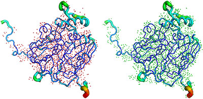
In a recent paper a group of scientists from France and the UK [Blakeley et al. (2015). IUCrJ. 2, 464-474; http://dx.doi.org/10.1107/S2052252515011239] outline the current rise in status of sub-atomic X-ray and neutron macromolecular crystallography and outline future prospects for combined approaches.
These trends are set to continue through further improvements planned to existing instrumentation, such as the addition of new detectors and the construction of entirely new instruments, such as the TOF Laue diffractometer “NMX” at the European Spallation Source (ESS). The researchers comment, however, that despite all the advances in the field, relative to X-rays, significantly larger crystals will always be required for neutron diffraction studies, particularly with the drive towards the study of ever-larger macromolecules and complexes. It is in this sense that further development of instrumentation and methods for large crystal growth are required. Creation of laboratories dedicated to the optimization of crystal volume and quality would help increase the effectiveness of neutron macromolecular crystallography. Significant efforts are required by the neutron community and facility providers to make neutron crystallography more accessible to the wider structural biology community.
Bill Stirling, Director of ILL said “While synchrotron X-ray methods dominate the field of macromolecular structure determination, neutron diffraction remains an extremely valuable complementary technique. As this article demonstrates, the power of the neutron technique resides in the ability to determine hydrogen positions, without radiation damage issues, allowing room temperature structure determinations. Further, new advances in instrumentation, exemplified by the LADI-III diffractometer at the ILL, exploit efficient Laue techniques that allow the use of much smaller crystals than previously possible and provide rapid and accurate structural information.
I have watched the development of neutron methods in biology over more than 40 years now – it is clear to me that Matthew Blakeley and his colleagues are providing biologists with extremely valuable, indeed essential, tools that will lead to major advances in our understanding of increasingly complex macromolecules."