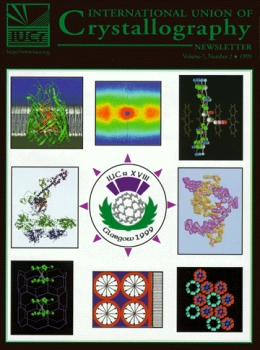
The full range of crystallographic research to be presented at the XVIII Congress of the International Union in Glasgow, August 4-13, 1999, is reflected in these illustrations (composite by Patricia Coley). Clockwise beginning at upper left: (1) Ribbon drawing of FepA, an active transporter of ferric enterobactin in the outer membrane of E. coli. S.K. Buchanan et al. (1999) Nature Structural Biology 6:56. Courtesy of Hans Deisenhofer. (2) Neutron quasi-Laue diffraction pattern from tetragonal hen egg-white lysozyme revealed 960 hydrogen atoms and 251 water molecules. N. Niimura et al., Nature Structural Biology, Vol.4, No. 11, 1997, pp 909-914. Courtesy of N. Niimura. (3) Anisotropic Displacement Parameters (ADP) of anthraquinone between 160 and 300K (Brock and Fu, Acta Cryst. B54 (1998) 308) shows butterfly-type motion of the molecule (S.C. Capelli, PhD thesis, U. of Bern, Switzerland, 1999). Picture by J. Hauser. Courtesy of H.-B Bürgi. (4) A 24-nucleotide RNA enzyme, Leadzyme, complexed with Sr(H2O)3(II) at 1.8 Å resolution reveals how small ribozymes recruit metals to activate specific 2'-hydroxyl groups for nucleophilic attack leading to phosphodiester scission. The substrate has a purple electron density surface and yellow nucleotide bonds. The ribozyme, a yellow electron density surface and pink nucleotide bonds. Courtesy of David McKay. (5) The hydrate of 18-crown-6/methylammonium fluoride, a channel structure in which water molecules form a 'double helix' along the crystallographic z axis. Courtesy of Janusz Lipkowski. (6) A model of a surfactant templated silicate structure at the air water interface of a cetyl trimethyl ammonium solution being studied by X-ray and neutron reflectivity. Courtesy of John White. (7) A very-large-pore high-silica zeolite UTD-1 bis(pentamethylcyclopentadienyl)cobalt(III) complex solved ab initio from powder diffraction data collected on a textured sample. Courtesy of Lynne B. McCusker. (8) The end of the myosin power stroke from crystal structure analysis of actin and myosin and electron microscopy of the actin-myosin complex. Courtesy of Ken Holmes.