
IUCr Congress
International Union of Crystallography
XVII Congress and General Assembly
Instrumentation and Experimental Techniques
In his keynote lecture, New Opportunities in X-ray Crystallography at Third Generation Synchrotron Sources, C. Branden reviewed recent achievements of the X-ray program at the European Synchrotron Radiation Facility (ESRF) in Grenoble. With cryocooling, macromolecular structures are routinely solved to 2.5 Å resolution from crystals with volumes as small as 104 μm3. Energy dispersive diffraction to 120 keV is used to follow in situ chemistry such as cement hydration where short lived intermediate phases have been found to have strong influence on the final state. Carbon monoxide release by photolysis from myoglobin has been followed on the nanosecond timescale. Using Laue diffraction, MAD experiments are routinely conducted on bending magnet beamlines equipped with a CCD. Beams 20 μm × 20 μm in size are used to obtain fiber patterns from 5 μm diameter spidersilk and capillary optics reduce the beamsize to 2 μm for diffraction and fluorescence mapping of small particles. Pressures equivalent to that at the center of the earth will be achievable in diamond anvil diffraction experiments and submicro beam sizes have been demonstrated with Bragg-Fresnel optics.
M. Hart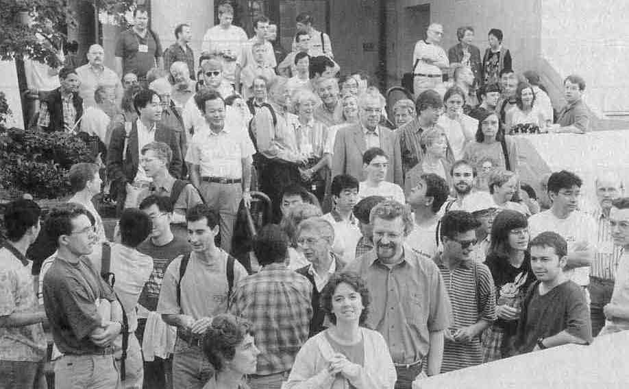 Queuing for the wine tour.
In the Microsymposium Synchrotron Radiation II - Macromolecules (1.03), M. E. Wall (Princeton U.) reported on the first complete digitization of a three-dimensional map of diffuse scattering from a crystal of a nuclease. He and his coworkers h ave analyzed this pattern of scattering to study the nature of the internal dynamics of the protein. Z. Dauter gave an overview of high-resolution macromolecular data collection (in the range 0.9-1.3 Å) as pioneered at the EMBL Outstation at Hamburg; detailed high-resolution refinements are now available for more than 20 protein structures, thus bridging the gap between small and large structures. The phase problem was central in four communications. E. Weckert (Karlrushe) reported on the phasing of tetragonal lysozyme using three-beam interference effects and W. Weis (Stanford U.) described the derivation of model-free protein phases by the MAD technique. The combination of the large anomalous scattering effect at the absorption edge of lanthanide ions and a large partial structure contribution from these ions led to phases of an unprecedented accuracy. M. Schiltz (LURE) discussed the use of xenon and krypton as heavy atoms and anomalous scatterers, and described a test experiment on an elastase crystal combining a SIRAS experiment and solvent flattening. Data were collected at a single wavelength on the same sample at normal pressure and under compressed krypton. The weak anomalous and isomorphous effects resulting from the binding of about 0.5 Kr atom per protein molecule were sufficient to get a very accurate electron density map. G. Privé (Toronto) described the phasing of a 450-atom peptide structure by the Shake-and-Bake direct method of structure determination. High resolution data obtained with SR at BNL's NSLS beamline X12-C was a prerequisite for this successful determination. Finally, C. Nave (Daresbury) elaborated on the optimization of data collection with SR: a timely communication indeed, since new instruments for macromolecular crystallography are being installed at several third generation SR facilities.
Roger Fourme and Robert Sweet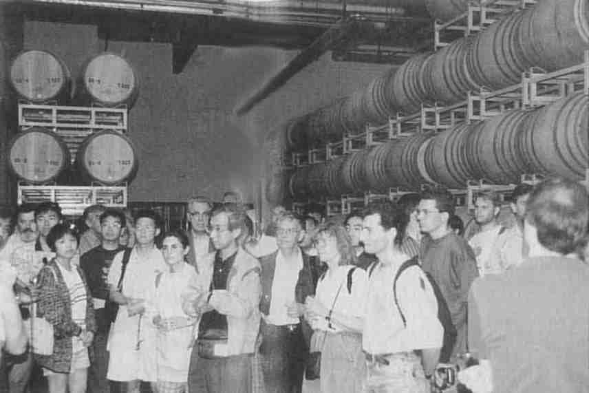 Barrels of fun on the excursion.
Detectors and Data Processing I: Macromolecular (1.04). The evolution of area detectors and data processing techniques for data acquisition in the last 20 years has been extensive. Film densitometry, gas detectors, TV systems, image plates (IPs), and CCDs have individual strengths and weaknesses. Multi-wire proportional counters, for example, have true counting accuracy and sensitivity but diminishing absorption and spatial resolution at short wavelengths. The use of absorption edge data from common metal atoms for phasing and high resolution analysis requires short wavelengths (e.g. 0.7 to 1.0 Å). Consequently, solid state detectors and higher energy synchrotron machines have been a principal focus of development. The strengths of CCDs and IPs include good sensitivity and preserved imaging capability with short wavelengths. Very impressive results have been obtained with on-line IP, very large IP (Weissenberg and Laue) off-line, and on-line CCD devices. It has become possible, using intense, tunable synchrotron radiation, to measure multiple wavelength data, to reach atomic resolution, and to record time-slicing dynamical protein crystallographic data. In chemical crystallography, area detectors in the home laboratory are beginning to make a major impact on precision and speed. For neutron crystallography IPs are beginning to be used in Japan (N. Niimura et al.) and the U.S. (C. Wilkinson, M. Lehmann et al.). Detectors with larger aperture IPs and better duty cycle CCDs are being sought. Tiling of CCDs into a 3 × 3 mosaic (E. Westbrook, APS) has recently been exploited. Increased count rate capability is expected with the "pixel detector," a silicon based device with independent pixel counting chains. N. H. Xuong presented results of a prototype device involving the recording of the direct beam into one of the counting chains. Ultimately a 1000 × 1000 pixels device will offer a major leap forward in detector performance. Pixel detectors development is also under way at San Diego/Berkeley (Xuong/T. Earnest), in Princeton (S. Gruner), and Imperial College (G. Hall). Pixel detectors can have a major impact in time-resolved experiments, MAD application, and in atomic resolution data collection in raising the molecular weight ceiling. With smaller protein crystals, in which absorption is less of a problem, longer wavelengths (e.g. ~ 1.5 Å) can be used for native and derivative data collection. At these wavelengths MWPCs can image without parallax problems, detector noise contributions are negligible, and count rates are manageable.
J. R. Helliwell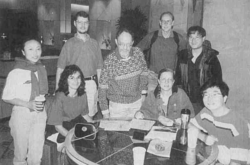 Ewald Prize winner, Michael Rossmann, and friends plan to take a hike.
Neutron Scattering I: Dynamical Aspects (1.07) The Neutron Scattering Commission attempted to cover the whole gamut of systems available to neutron investigation from electronic excitations in magnetic systems to the complexity of slow motions in biomolecular aggregates. No casualties were reported! Energy levels from microelectron volts to electron volts can be studied using inelastic neutron scattering and the intensity of the modes is directly related to the amplitude of vibration of the atom involved. After hearing of the latest studies of the dynamics of high T's (M. Arai) we moved to the rotational dynamics of ammonium ions (S. Belushkin). J. Eckert showed data on the dynamics of hydrogen in coordination complexes and J. Larese demonstrated the sensitivity to quantum tunnelling of molecules on surfaces as you move from two-dimensional to three-dimensional behavior. B. Asmissan reviewed beautifully systematic work on rotational dynamics of methane in rare gases and its theoretical interpretation. Finally A. Deriu shared latest results on the study of the diffusion of water in bio-gels.
A. AlbinatiThe session on Electron Diffraction from Surfaces (1.08) emphasized the diversity of approaches to surface structure analysis using various forms of electron diffraction. Some of the techniques resemble X-ray diffraction in that the source of diffracting electrons is located far from the sample, giving rise to incident plane waves. This applies to low-energy electron diffraction (LEED), the oldest and most productive technique for surface structure determination described by K. A. R. Mitchell. It also applies to reflection high-energy electron diffraction (RHEED), which is rapidly developing into a powerful competing technique, described by A. Ichimiya. For a novel point reflection microscope (J. Spence), the source of electrons, a field-emitter tip, is located within hundreds of ångstroms of the surface: it produces a diverging electron beam, multiplied into several divergent diffracted beams which produce images carrying three-dimensional structural information. Other techniques use point sources of electrons that are located at atomic sites of the surface itself. Thus, photo- and Auger-electron diffraction, discussed by S. A. Chambers, both utilize the diffraction of electrons by atoms surrounding the emitting atoms to collect structural information. These two techniques allow energy discrimination of electrons coming from different elements, or from the same element in different chemical environments. Another point source technique, which lacks chemical discrimination, is Kikuchi electron diffraction, described by C.-M. Wei. The three techniques were also presented in their "holographic" form: the diffraction patterns can be viewed as holograms, which often can be inverted by suitable Fourier transformation to produce real-space three-dimensional images of local surface structures. These images are invaluable as starting points for more refined structural determination using trial-and-error searches. The effects of multiple scattering and the twin image problem on inversion schemes were discussed by several speakers, and emerged as crucial theoretical issues.
Michel Van HoveMethods of Structure Determination
In his keynote address, Mad Phasing Theory and Practice, W. Hendrickson summarized the development and current status of multiwavelength anomalous diffraction (MAD) phasing of X-ray data from macromolecular crystals. The potential of anomalous scattering has long been recognized, but it is the availability of readily tunable synchrotron sources that has led to the explosive growth in the number of macromolecular structures solved by the MAD procedure in recent years. Choice of suitable wavelengths for collecting data enables the investigator to exploit the relatively large changes in both the real and imaginary components of the atomic scattering factors at energies near the binding energy of the inner electrons of the heavier atoms (Z greater than 20). Such elements are present in some proteins, and can be introduced into others. For example Se can replace S in methionine which can then be incorporated during protein synthesis. Atoms in the lanthanide series of elements exhibit particularly strong anomalous scattering and substituted for Ca in a protein have proved very effective in MAD phasing. The method is becoming in many cases the method of choice for ab initio phasing of X-ray data from macromolecular crystals.
Lyle Jensen Not really naked!
 Point Man: Ron Stenkamp.
The focus of the Microsymposium Direct Methods of Phase Determination (2.03) summarized by F. Hai-fu, was the transition of direct methods application to problems outside of their traditional areas from small to large molecules, single to powder crystals, periodic to incommensurate structures, and from X-ray to electron diffraction data. I. Karle opened the session with useful advice based upon her applications in the peptide field. Highlights in the session included the description by W. Lasocha of successful application of direct methods to powder data and the talk by R. Miller on the Shake-and-Bake approach, used in the determination of several large small molecules and proteins. Talks on new direct methods and applications were presented in other Microsymposia reflecting growth in the field.
Suzanne FortierComputing in Modeling Analysis and Design
The Internet (3.01) pervades all aspects of everyday professional and private life. The WWW has fundamentally altered information delivery. The Microsymposium on the topic reviewed recent experiences, advances, and technical developments significant to crystallography. The chairperson of the Electronic Publishing Committee of the IUCr described the advantages, disadvantages, and cost of electronic versus conventional publishing. In particular the difficulties with electronic publishing were detailed in relation to the requirements for IUCr journals. A strategy involving active participation by relevant IUCr Commissions was considered essential, and one practical scenario was detailed. It is always interesting to observe how revolutions in technology influence the working style of scientists and, consequently, the overview of the role of the World Wide Web in computational and pharmaceutical chemistry provided food for thought. Running scientific conferences entirely electronically over the Internet is one way to diversify with cost savings. A survey of databases, information and teaching sources, experience in electronic publishing, and a Crystallographer's Guide to Internet Tools and Resources were presented. The Java programming language and the virtual reality modelling language (VRML) are certain to have an impact on Web use by crystallographers. Both languages were demonstrated in applications of interactive graphical representations of molecules and crystals, and the advantages and disadvantages described. The session chairman was H. D. Flack, the vice-chairman, Y. Epelboin and the speakers were E. N. Maslen, G. D. Purvis, Y. Epelboin, J. C. Huffman and A. LeBail.
Howard Flack The Scottish cultural attaché and his Epicurean advisers plan the haggis fest for the Glasgow banquet.
An Internet Workshop. A group of enthusiastic tutors gave brief presentations of their area of Internet knowledge and experience. This was followed by hands-on demonstrations using UNIX workstations, PCs and MACs. The organizer compiled a hypertext documentation series of World Wide Web files called "A Crystallographer's Guide to Internet Tools and Resources" complete with a table of contents and indices. The W3 is being increasingly used as a convenient graphics and distributed front end to programs installed on a server. The Internet has been used in the organization of crystallographic conferences, for the distribution of information on Crystallographic associations, for access to the World Directory of Crystallogaphers, IUCr journal submission, and by use of News-groups (sci.technique.xtallography and bionet.xtallography). A Crystallographer's Guide to Internet Tools and Resources is freely available over the WWW at URL: http://www.unige.ch/crystal/w3vlc/int.index.html.
Howard Flack Water games at the Seattle Science Center.
Macromolecular Map Fitting and Modification (3.03) began with a brief overview (K. Cowtan) describing the current status of density modification methods, their successes and limitations, and the convergence between the map modification and map fitting disciplines. K. Das gave an example of electron density averaging between crystal forms in the structure determination of HIV1-RT. His innovations included local scaling of the related densities, averaging omit maps to reduce bias, and the use of a reciprocal space convergence criterion. These techniques combine to reduce the problems of bias to an initial structural model. D. Turk described his "Main" package, which combines model building, refinement, and density modification in an integrated manner. The continuous consultation of the experimental observations allowed by this method facilitates structure building and reduces errors. C. Carter reported a difficult structure solution using molecular replacement in which bias to the starting model was a serious problem. His approach combined non-crystallographic symmetry averaging using maximum entropy maps, and phase permutation using likelihood ranking. A. Szoke described a new method of combining real and reciprocal space information through a wavelet-related technique which he describes as a holographic method. Some test calculations using his program, "Eden", demonstrate the power of this method to solve a structure from a very small known fragment. This was widely commented on as the most exciting talk of the session. D. McRee described the latest facilities for map fitting in XtalView, including automated fitting of main chain and side chain residues. T. Oldfield went on to talk about his work towards the semi-automation of the map fitting process, including methods for automatically identifying secondary structure features in the map, connecting these features, fitting the sequence, and then fitting and refining the main and side chains. His knowledge based approach, implemented in Quanta, builds on all the information available in the PDB from solved structures.
Kevin CowtanThe Materials Research (3.06) session illustrated how effectively molecular simulation can contribute to crystal structure determination and analysis. Simulated annealing was presented as a means of circumventing the phase problem by allowing direct refinement of initially random models. C. M. Freeman's overview described compelling applications of this direct space structure solution route to both inorganic and molecular systems. To accelerate model-building approaches or as an aid in rationalizing structures from simulation, Y. Le Page and C. M. Kölmel presented methods to determine space group symmetry from initially triclinic descriptions; fully-automatic ways to derive unit cells, space group symmetries, and asymmetric unit descriptions. Other talks concerned prediction of molecular crystal structures (M. U. Schmidt), use of periodic Hartree-Fock calculations to rationalize the complex behavior of ostensibly simple oxides and yield a range of computed properties (N. Harrison), and simulation of the geometries and energetics of point defects in oxides (R. A. Jackson).
J. M. Newsam (Front row) Anders Liljas, Ellie Adman, Kathryn Ely; (back row) Chrishan Riekel, Toshihiko Takama, Yoshihito Nakane, Francoise Riekel, and Guenther Eulenberger.
Biological Macromolecules
In his keynote address, The Structure of Bovine Mitochondrial F1-ATPase - An Example of Rotational Catalysis, A. G. W. Leslie described a 350 kD assembly responsible for the synthesis of ATP consisting of 3 α/β dimers, and a single γ subunit at the center of the assembly. In cells the F1-ATPase is complexed to a transmembrane gated proton channel (Fo) that provides the driving force for the synthesis of ATP. The structure of ATPase is the first crystallographic analysis to reveal how chemical and mechanical transformations are coupled in proteins. Leslie showed us that the three different α/β pairs of subunits, although identical in sequence, assume different conformations in the assembly. Apparently, the active sites of the F1-ATPase alternatively assume conformations complementary to substrates, complementary to product, and open, as a function of the position of the γ subunit. The structure beautifully validates the conclusions about the action of the F1-ATPase based on biochemical studies. The rotation of the γ subunit, which causes the subunit holding ADP + Pi to change into a conformation complementary to ATP, is thought to occur through the action of the proton pump, by a mechanism yet to be elucidated.
Elizabeth GoldsmithIn his keynote address, The Integration of Structure-Based Drug Design and Combinatorial Chemistry for Efficient Drug Design, R. Salemme described some of the major challenges and problems encountered in structure-based drug design, and ways that combinatorial chemistry can expedite the discovery of lead compounds that are candidates for iterative improvement. Salemme reviewed a series of thermodynamic and structural studies of ligand binding to strepavidin, which binds biotin and analogs of biotin with extremely high affinities. These studies clearly show the challenging difficulties of rationalizing and predicting binding constants. Since the free energy of binding includes both enthalpic and entropic components, subtle variations in interactions between ligands and the protein, coupled with changes in solvent structure and conformational freedom, can produce large and unpredictable changes in binding constants. The predictive power of current modelling approaches is quite limited, even when detailed structural data are available. Consequently, structure-based drug design proves to be most powerful when used in an iterative manner to improve the properties of lead compounds. One method now being used for identifying leads involves automated screening of compound libraries produced by combinatorial chemistry. Salemme described a novel combinatorial approach for developing libraries of lead compounds to block the active site of thrombin, a serine protease involved in blood clotting. Thrombin has three major subsites within the active site of the enzyme. By combining multiple chemical groupings that might interact with one or more of these three subsites, he has produced lead candidates that were then optimized by structure-based techniques to produce potent and selective inhibitors of thrombin. One of these thrombin inhibitors will soon be advanced into clinical trials.
Charles Bugg Speakers in the Microsymposium on MAD and EXAFS. Front row (left to right): N. Blackburn, J. Penner-Hahn, P. Lindley; back row: C. Kratky, K. Hodgson, P. Evans.
Keynote Lecturer J. Thornton, Protein Conformational Analysis, focused on developing approaches and software tools that facilitate automated validation of protein structures and aid in their analysis and classification. Methods for validating structures that are based on knowledge derived from known structures were discussed, as was the effect of including information from recently solved, atomic-resolution protein structures. A hierarchical system was presented that describes protein structures in terms of class, architecture, topology, and homologous superfamilies. It was clear that as the number of known protein structures grows, our need to analyze properly these structures will increase dramatically if we are to succeed in mining the wealth of information to be found in our structural databases.
Judith Kelly Marcia Vair wonders "Do l dare to eat a wing" as Bob Bryan, Ciel Steinfink, and Patti Coley look on.
The Microsymposium on Enzymes (4.01) can be summed up under the themes of unity and diversity. Unity in the strategic importance of structure for understanding mechanism, first exemplified 30 years ago with lysozyme, as G. Petsko reminded us, and diversity in the vast number of enzymes and substrates available for study. D. Barford's talk on protein phosphatase demonstrated beautifully the advances in understanding the mechanisms of tyrosine phosphatase. E. Goldsmith followed with an account of the regulatory properties on MAP kinase. Moving from proteins to DNA as substrate, X. Cheng's talk on DNA methyl transferase provided an example of substrate modification before catalysis. In a base flipping mechanism, the enzyme takes the base to be modified into the catalytic site and replaces the vacant site on the DNA with a loop from the protein. B. Dijkstra completed the analysis of proteins that act on macromolecules with a description of muramidase, the extraordinary all α-helical doughnut-shaped protein that catalyses the hydrolysis/transglycosylation reaction. This chemically attractive mechanism had been considered for lysozyme but ruled out on structural and chemical evidence. It is interesting to see that other enzymes can exploit it.
Turning to enzymes that act on small substrates, C. Hasemann described the recently determined structure of the bifunctional enzyme, 6-phosphofructo-2-kinase/fructose 2,6 bisphosphatase which showed that the kinase domain resembled adenylate kinase and the phosphatase domain phosphoglycerate mutase and that the catalytic sites on the two domains were well separated. The final two talks illustrated some unexpected themes. The structure of urate oxidase, described by N. Colloc'h, exhibits a 16 strand β barrel in which each of the four subunits contributes four strands. The catalytic site, identified from inhibitor binding studies, shows no metals, no cofactor, and no functional amino acids. The enzyme appears to act by correctly locating the two substrates, urate and oxygen. L. Howell ended the session with a demonstration of the significance of structure for mechanism in the studies on arginosuccinate lyasel/δ crystallin. Genetic studies had shown that residues important for catalysis are located on parts of the chain far apart in sequence. In the structure these are far apart in space on the subunit but brought together in the tetrameric assembly, thus providing an explanation for intragenic complementarity, a mechanism in which an active multimeric protein can be assembled from monomers produced by two different inactive mutant alleles of the same gene.
Louise JohnsonMetalloenzymes (4.02). The past few years have seen a flowering of the field with the first structures of vanadium, nickel, cobalt, and tungsten-dependent enzymes; the spectacular detail of the multi-metal cytochrome oxidase structure; and the strange twists of metalloenzymes obtained from archaebacteria. E. Jabri described the recently determined structures of the nickel enzyme urease, the first enzyme ever to be crystallized, which was not known to contain nickel until many years later. She showed how its unusual metal geometry is exquisitely defined by its surroundings. Other presentations ranged through enzymes dependent on iron [sulfite reductase (B. Crane), and a PCB degrading dioxygenase (Y. Mitsui)], copper [nitrate reductase (E. Adman)] and zinc [glyoxalase I (A. Cameron)]. Perhaps the highlight, however, were the molybdenum-dependent enzymes of the two final presentations, by J. Boyington and H. Schindelin. The two examples illustrated special properties of the molybdoenzymes including unusual chemistry and ligands. Most spectacular of all, H. Schindelin finished the session with the first view of the complete nitrogenase complex, comprising both the Fe/Mo and Fe proteins. The trick to obtaining a stable, crystallizable complex seemed to have been the use of ADP and AlF
 A roundup of the usual suspects in the Protein-RNA imbroglio. Front row: K. Nagai, M. Safro, D. Tomchick, S. Cusack (formerly S. Nikonov, Vol. 4, No.2, p. 9), J. Nyborg; back row: C. Romiert, T. Steitz, D. Moras.
The Protein-DNA Session (4.05) featured researchers tackling the problem of understanding how proteins recognize and modify DNA from many different vantage points. C. Calladine (Cambridge U.) presented results of detailed analyses of DNA conformations in protein-DNA complexes and demonstrated the structural changes in DNA caused by binding of the catabolite activator protein. M. Lewis (U. of Pennsylvania) and J. Geiger (Yale U.) described cocrystal structures in which protein binding causes DNA deformation. The Lac repressor operator complex (Lewis) revealed a pair of alpha helices binding in the minor groove creating a bent DNA stucture that provides a model for binding of Lac repressor to all three operators of a canonical Lac operon. Geiger presented the structure of a triple complex of transcription factor IIA, a TATA box-binding protein and a TATA element. In sharp contrast to the Penn work, this structure demonstrates minor groove binding and DNA bending by the anti-parallel β sheet of the TATA box-binding protein. In the arena of molecules that perform chemistry on DNA, talks were presented by D. Vassylyev (Protein Engineering Res. Inst.), S. Fujii (Osaka U.), A. Mondragon (Northwestern U.), and L. Beese (Duke U.). Vassylyev and Fujii described molecules that participate in DNA repair, with the PERI structure demonstrating base flipping. Mondragon's topoisomerase structure suggested an intriguing model for its mechanism of action. The high point of the session was an even more penetrating view of a similarly complicated enzyme. Beese presented the structure of a thermostable DNA polymerase at work! She has been able to record diffraction data from a cocrystal of the polymerase with substrate, add a single nucleotide triphosphate to the crystal and then repeat the crystallographic experiment. The resulting data gave a Fourier synthesis that showed that the polymerase had catalyzed single base addition to the substrate within the confines of the crystal lattice. Technical advances in computing, X-ray production, position-sensitive X-ray detectors, cryopreservation of the crystal, and multiwavelength anomalous dispersion are allowing us to tackle ever larger and more difficult problems.
Stephen K. BurleyThe ten Hot Macromolecular Structures I (4.07a) selected for oral presentation included intracellular regulatory molecules, extracellular recognition molecules, and molecules of medical importance in response pathways.
The structure of the 26 nucleotide RNA pseudoknot inhibitor of reverse transcriptase described by C. Kundrot showed that nucleic acids as well as proteins have an ability for domain swapping. The two intracellular regulatory proteins described exhibited all helical topologies. Cyclin H, a component of the cell cycle activating kinase (CAK) presented by K. Kim, has a similar structure to cyclin A, despite only 12% sequence identity, but sufficient differences to indicate that its interactions with CDK7 are different from those of cyclin A with CDK2. Bcl-XL, described by S. Muchmore, is a component of a family of proteins that modulate apoptosis. The helical fold in the structure is similar to that in diphtheria toxin and the globular domain of colicin, suggestive of a role for this protein in transmembrane events, but the structure also contains a 43 amino acid loop with possible involvement in control by phosphorylation. The T cell membrane receptor described by C. Garcia has the familiar immunoglobulin fold for the variable and constant domains but with sufficient differences from the standard fold that the structure could only be solved by MIR.
 Did anyone else get stoned in Seattle?
The structure of colicin A, reported by M. Wiener, demonstrated how we can still be delighted and surprised by new structures. The channel forming bacterial toxin has three distinct domains involved in receptor binding, translocation, and pore formation linked by an extraordinarily long (160 Å) helical coiled coil. The crystals were 80% solvent and the structure had been solved by engineering cysteine residues. It seemed remarkable that such a non-globular structure had crystallized. Equally surprising was V. Malashkevich's structure of the pentameric glycoprotein heptad repeat domain from the cartilage oligomeric matrix protein showed five α helices 73 Å in length wrapped around each other to form a superhelix with a hydrophobic channel in the center, a polar exterior, and a chloride ion in the center. On exposure to xenon gas, eight xenon atoms were located at defined sites in the channel. This is the first example of a five helix bundle at atomic resolution and has significant implications for the structures of ion channel proteins such as the nicotinic acetylcholine receptor. T. Greenough described the five fold symmetric C reactive protein involved in acute phase response. This speaker remained remarkably cool despite problems with the projector that resulted in his last six slides being placed in random order. V. Lee described the structure of a 30K C-terminal fragment of γ fibrinogen.
The human lysosomal sulphatase, described by C. Bond, is an enzyme involved in processing N-galactosamine-4-sulphate. The structure, the first example of this class of enzymes, showed two domains, one of which, with ten strands of β sheet, resembled unexpectedly the structure of alkaline phosphatase. The final structure of proteinase involved in apoptosis, described by J. Rotonda, showed similarities to Interleukin-1β Converting Enzyme but with differences in inhibitor binding subsites that will certainly be of interest to the pharmaceutical industry intent on manipulating this biological pathway.
Louise Johnson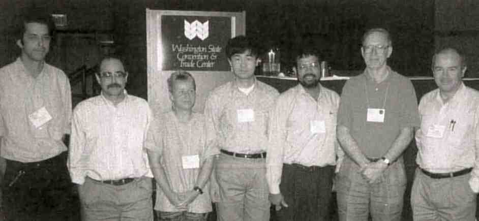 Speakers in the Membrane Protein session doing an impression of lipid lipid association (left to right): T. Schirmer, M. Garavito, S. Karrasch, D. Xia, G. Kurumbail, N. Isaac and J. Deisenhofer.
Macromolecular Assemblies (4.08). The session started with a description of the pyruvate dehydrogenase complex of which the icosahedral core had just been solved at 5 Å resolution by T. Izard. The outer diameter of the 60-meric particle is 230 Å, the inner diameter is 140 Å and there is a large pore of ~55 Å cross section, which allow CoA to diffuse inside and find the acetyl group bound to lipoyl domains which swing around the core in a dynamic fashion.
S. Burley described the fascinating complex of the "TATA Box" bound to the TATA Box binding protein (TBP) and the Transcription factor TFIIB. In the complex, crucial for the initiation of transcription in all living organisms, the TBP-TATA binary complex has virtually the same structure as in the TBP-TATA-TFIIA ternary complex. Together these structures suggest a framework for future experiments and speculations concerning such things as the role of TATA box-binding protein associated factors (TAFs) whose structures were also reported.
A. Yonath described continuing efforts to reveal the structure of the ribosome. A series of poly-heavy atom derivatives have produced new electron density maps at low resolution. The goal of obtaining better ribosome crystals via new purification methods was rewarded by crystals diffracting to 1.9 Å. These turned out, however, to be of "degradosome" multi-protein complexes of 250,000 dalton associated with ribosomes. Although not the ultimate goal still a most remarkable achievement.
S. Sprang described the intricate structure of the trimeric G protein Giαβγ. Solved by a combination of MR, MAD and MIR, the catalytic mechanism regions responsible for important conformational changes in the γ-component were identified. The β subunit contained seven propeller motifs plus a number of important helices. The γ subunit is much smaller and only interacts with β, not with α. The γ subunit has numerous interactions with β in such a manner that it stabilizes the GDP form of Giα. Mapping biochemical data onto the structure is providing a rapidly increasing degree of insight.
P. Alzari described the organization of the wonderful cellusome which provides "up to nine hydrolytic enzymes on a string" outside the cell- all attacking cellulose. The scaffolding protein is CipA which has a "dockerin domain" that allows it to bind to the outer membrane. It contains nine dockerin receptor domains, each of which can bind a hydrolytic enzyme which all have a dockerin domain at the C-terminus. The set of enzymes on a string has no constant stoichiometry but an apparent evolutionary advantage.
J. Tainer described the structure of a pilin dimer in which two subunits are interacting via an anti-parallel coiled coil, burying 1100 Å2 of surface. This interaction is considered to be a crystallization artifact and the organization of the pilin in the fiber is thought to be quite different, with five subunits per turn, a pitch of 41 Å, and carbohydrates and hyper-variable regions on the outside. A video illustrated dramatically the wonder of the assembly process.
P. Sigler described the structure and action of GroEL which, alone or in complex with GroES, prevents other proteins from miss folding. The mechanism of action is still not completely settled, so, instead of using sexy Southern California style slides, Paul showed schematic transparencies. Otherwise the audience might believe that the proposed mechanism was real. The structure determination of the double ring of seven subunits in the tetradecameric GroEL cylinder was truly a remarkable feat, as was its refinement by the Brunger group using torsional molecular dynamics procedures.
Wim Hol Program chair and support staff: C. Zardecki, S. Duax, J. Duax, T. O'Conner, H. Nova de Armas, M. Sawicki, A. DeCecco, A. Haake, N. Li.
Muscle and Motor Proteins (4.13). In the past year great advances have been made in the understanding of the molecular motor, myosin. C. Smith reported on the recently published structure of a fragment of myosin cross bridge crystallized with ADP vanadate as an analog of the transition state complex. This form would be expected to show the beginning of the power stroke in muscle and indeed it does. Equally exciting was the report from R. Milligan on high resolution cryo electron microscopy of actin decoated with myosin cross bridges, where on addition of ADP he was able to see the distal lever arm of the myosin cross bridge rotate through 35°. The crystal structures of two forms of a second molecule motor, kinesin, reported by J. Kull and E. Sablin, demonstrated an unexpected similarity with myosin and showed evidence of a common mechanism, a mechanism similar to that of the G-proteins. Burtnik described the structure of gelsolin, an actin severing protein, which has six similar sub domains and is capable of a rich polymorphism.
R. Fletterick The Keynote addresses were so well attended that you had to get there early to be in the second row.
The Symposium Macromolecule Based Drug Design (4.12) provided a forum to examine how structure-based design is done in industry and to call attention to challenges and opportunities in the field. The commonly recognized first structure-based drug is the angiotensin-converting enzyme (ACE) inhibitor Captopril. The structure of carboxypeptidase A, a zinc-dependent exopeptidase, provided the structural underpinnings for the design process based on the hypothesis that ACE, a zinc-dependent exodipeptidase, worked like carboxy-peptidase A. In the subsequent decade, the concepts of structure-based drug design were developed, refined, and popularized in a number of visionary labs around the world. This decade marked the birth of the biotechnology industry, a growing realization of the potential of structure-based design, and the first wave of additions of crystallographic groups to pharmaceutical research organizations, which continued into the late 80s.
The subject of the first presentation was Trusopt, a carbonic anhydrase inhibitor used to treat glaucoma. Presentations on HIV protease and thrombin illustrated the evolution of structure-based drug design in the last decade. Other presentations concerned HIV reverse transcriptase, influenza virus neuraminidase, human renin and plasmepsin, and the causative agent for malaria. One talk opened discussion in a new area of design, small-molecule antagonists of protein-protein interactions. There is prejudice against the idea that a small molecule can prevent, attenuate, or alter the binding of a macromolecular ligand to its receptor, or the formation of function in hetero-oligomeric complexes. However, examples such as HSV ribonucleotide reductase, where a peptidomimetic of the C-terminus of one subunit inhibits subunit association, and FGF, where poly-saccharide mimics of heparin enhance the binding of ligand to receptor, indicate that it can be done. The example of the calcineurin-FKBP12-FK506 complex illustrated how the drug FK5 06 facilitates protein-protein interaction between calcineurin and its binding protein. Such structural findings suggest new strategies for design of novel analogs to alter protein-protein interaction.
The picture of how drug design is currently pursued in industry includes the following observations: Placement and Support. Integration of the structure determination group for ready access to protein samples is critically important. Role of Chemistry. Chemistry programs have a momentum of their own and create many more compounds than can be examined crystallographically in a useful time period. The key to structure-based design in these circumstances is to determine which compounds NOT to study and here computational chemists are of great help. Two kinds of compounds are of special interest: new chemical leads and those molecules that inexplicably violate current structure-activity relationships (SAR). Structure-Based Drug Development. Other properties besides biological activity, such as solubility and ease of uptake, ability to reach the appropriate site of action in vivo, resistance to degradation, and avoidance of rapid excretion are desirable. Some of these properties can now be understood and manipulated at the molecular level and can thus be the subjects of structure-based design, too.
M. Grütter and H. Einspahr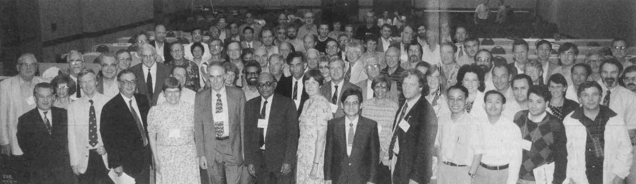
The delegates to the General Assembly, Executive Commitlee members in the front row: M. Hart, T. Baker (President 1996-1999), A. Authier, S. Larsen, P. Coppens (President 1993-1996), R. Chidambaram, P. Codding, J. Harada and H. Schenk. Robert Bryan, the chair of the meeting with a relieved smile on his face, is immediately behind P. Codding.
