Commentary
Learning about CSD-Particle at the ECM33 CCDC Workshop
![[Fig. 1]](https://www.iucr.org/__data/assets/image/0018/155304/Fig.-1.png "Fig. 1")
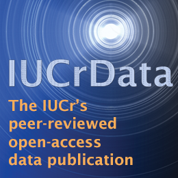
Figure 1. Visualising roughness from the topology of the (0 -1 -1) surface of theophylline form II (BAPLOT01). © CCDC.
I arrived a day early at the 33rd European Crystallographic Meeting (ECM33) to learn how to get the most out of chemical, materials and biological structures using the latest developments made by the Cambridge Crystallographic Data Centre (CCDC) at their day-long workshop. This was in the University of Versailles, approximately a half-hour walk from the centre of Versailles. To my knowledge, this was the first time that the CCDC had included biological structures as a topic in their educational workshops. This involved learning about the biomolecular graphics program Hermes to accompany their chemical crystal structures program of long-standing, Mercury. A new product suite within Mercury called CSD-Particle was also demonstrated, enabling the rapid analysis of mechanical and chemical properties of crystalline particles. This new software tool grabbed my attention. I am not surprised it is described in news from the Cambridge Structural Database (CSD) in this issue of the IUCr Newsletter. My short article is not a product review (for such see e.g. ref [1]); rather, I describe the aspects that impressed me.
CSD-Particle uses computational models and algorithms, with the data from the CSD, to provide both qualitative and quantitative analyses of particle shape as well as surface and bulk properties. It provides new insights into the properties of materials. As crystallographers and structural chemists, we are very familiar with calculating, for example, the optical properties of crystals or the Mohs scale of the hardness of materials such as by Lawrence Bragg, as he describes in his book [2]. For the former, Bragg calculated the refractive indices of calcite for light travelling in the planar carbonate groups and perpendicular to them, thus quantitatively explaining the double refraction effect. For the latter, he considered the extreme examples of soft graphite or talc, with weak bonding between layers of atoms allowing the layers to slide, versus diamond with its isotropic compressibility. Also, the electrical properties of metals, semiconductors and insulators arise from the electrons in crystals arranged in so-called energy bands [3].
CSD-Particle offers calculations of the potential chemical interactions at a particle’s surface, giving insights into its wettability, tabletability, flow and sticking potential. It also generalises the mechanical properties of a crystal by calculating its likely slip planes, related, of course, to the simple examples mentioned above. By quantification of the density of hydrogen-bond donors, acceptors, aromatic bonds, unsatisfied hydrogen-bond donors, the root mean standard deviation of the surface height, surface area, rugosity (see Fig. 1), kurtosis and skewness, CSD-Particle opens doors to molecular design.
I wonder if these could be applied to protein–ligand, surface to surface, interactions and the challenge of binding energy estimations [see 4]. For the latter, as emphasised by [4], the extension of static crystal structures to include molecular dynamics was necessary for a better quantitative calculation of a protein saccharide binding energy. To add experimentally determined hydrogen-atom positions using modern neutron protein crystallography would be an improvement on the X-ray only crystal structures used in [4].
The name ‘particle’ to me doesn’t quite fit the bill. As a trained physicist, it is a word reserved for ‘waves and particles’ and the ‘constituents of the atom’. To an environmentalist, its polluting particles are maybe closer to the CSD’s particle definition. In a lateral thought, and experiencing tea made in France at a café close to the ECM33 Congress Centre, I reflected on the optimum size of tea leaves. If too large, the poor surface area to volume ratio leads to too lengthy a time being needed to brew, and one’s impatience means a cup that is basically hot water and milk. For sure, though, the relationship of size, morphology, surface chemistry and function are closely related.
All in all, this new product, CSD-Particle, opens numerous new applied research directions in the whole topic of structure and function.
Acknowledgements
I am very grateful to the CSD staff who hosted the training event at ECM33, namely Ilaria Gimondi, Suzanna Ward and Natalie Johnson, as well as Arie van der Lee and the local organising team, who were a great help in organising the workshop. They all did an excellent job.
References
[1] Albert, H. (2022). Software from Cambridge crystallographic experts could save pharma industry millions. Chem. World. 4016026.
[2] Bragg, W. L. (1975). The Development of X-ray Analysis, pp. 166, 170. London: G. Bell and Sons.
[3] Kittel, C. (1976). Introduction to Solid State Physics, 5th ed., chapter 7. New York: John Wiley & Sons.
[4] Bradbrook, G. M., Gleichmann, T., Harrop, S. J., Habash, J., Raftery, J., Kalb (Gilboa), A. J., Yariv, J., Hillier, I. H. & Helliwell, J. R. (1998). X–ray and molecular dynamics studies of concanavalin A glucoside and mannoside complexes: Relating structure to thermodynamics of binding. Faraday Trans. 94(11), 1603–1611.
John R. Helliwell is Emeritus Professor of Chemistry at the University of Manchester, UK.
Copyright © - All Rights Reserved - International Union of Crystallography
The permanent URL for this article is https://www.iucr.org/news/newsletter/volume-30/number-3/learning-about-csd-particle-at-the-ecm33-ccdc-workshop