
Meeting report
Neutrons in Biology
A Neutrons in Biology Conference was held in Santa Fe in October 1994. The conference commenced with a welcome from J. Browne (LANL). In a keynote address entitled 'Neutron Anatomy', G. Bacon (U. Sheffield) described fascinating work on the orientation of polycrysta1line aggregates in bones. The details of the preferred orientation of crystals of hexagonal hydroxyapatite determine the ability of the material to withstand stress in any direction. There is a difference in the orientation of the hydroxyapatite in bone between human infants and lambs. Neutron diffraction orientation diagrams of the human tibiae have also been used to identify the differences in the lifestyle of two Neolithic tribes (cliff-dwellers and flatland dwellers) and modern sedentary tribes (neutron scatterers for example).
The discussion on the relative merits of steady state and pulsed neutron sources continues. D. Price (ANL) commenced with an overview of the present situation and J. Rush (NIST) continued to astound us with the ability to extract more neutrons from their reactor source and more dollars from the treasury. The long and short pulse story from R. Pynn (LANL) highlighted the emergence of pulsed neutron sources as major contributors to the field. Such was the enthusiasm displayed by C. Wilson (RAL) for the potential for biological crystallography and pulsed neutrons, that he clearly won the award for the largest number of words delivered in a 30 minute paper.
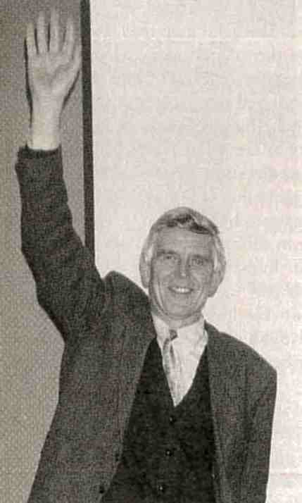 Heinrich Stuhrmann (i) asking to leave the room, (ii) demonstrating how big he would like next year's budget to be or, (iii) pretending to be a polarized neutron about to interact with a biologically important proton. (Photo courtesy of R. Knott.)
The second session on instrumentation highlighted the eternal struggle for more neutrons and more efficient instruments with presentations from diffractometrists at four major facilities, BNL (Y. Radeka and R. Korszun), ANSTO (R. Knott), ILL (S. Mason) and JAERI (N. Niimura and M. Imai). V. Radeka described the slow steady progress on the development of high precision thermal neutron position sensitive detectors and mentioned the astounding performance that can now be achieved. R. Knott surveyed neutron scattering Down Under and the prospects for a bright future with a proposal for a new neutron source. R. Korszun described the three stations at BNL dedicated to biological structure determination. The recent developments, particularly the new optics on the SANS instrument, were highlighted. News of competing priorities from non-biological applications and encouragement on the future of the ILL reactor came from S. Mason. N. Niimura presented a general overview of both reactor and pulsed neutron sources in Japan, and M. Imai completed this section with more detail on the SANS instruments on the JRR-3M reactor.
In the SANS session, V. Ramakrishnan (BNL) unraveled some of the mysteries of chromatin structure. B. Medelson (UC) showed how muscles are now better understood. G. Olah (LANL) presented a Monte Carlo modeling analysis. D. Svergun (EMBL) applied SANS analysis to the 50S ribosomal subunit from E. coli, and R. Hjelm (LANL) had the gall to probe the self assembly in mixed aqueous colloids of bile salts and phosphatidylcholine.
In the session on Membrane Structures and Dynamics, J. Bradshaw (U. of Edinburgh) probed the interaction of amphipathetic helices with phospholipid bilayers, S. White (UC) looked closely at the membrane data in "composition space" and S. Krueger (NIST) reflected on the structure of a single lipid bilayer. D. Worcester (UM) continued his methodical investigation of the structure of lipid bilayer systems and some fascinating studies of cylindrical aggregates of chlorophylls, and D. Middendorf (Oxford U.) completed the session with a comprehensive survey of the status of mode coupling and H-bond dynamics in biomolecules using pulsed neutron techniques.
A superb conference dinner created an atmosphere ideally suited for the discussion of the politics of science, and the idiosyncrasies of neutron scattering. In his after dinner speech, S. White implored the community to unify its position on the future of the major neutron scattering facilities in the US.
In a session on protein structure, P. Timmins (ILL) described using low resolution crystallography on membrane bound proteins. Through the use of contrast variation, the interactions of detergents with proteins are being investigated using single crystals about the same size as those used for X-ray diffraction. The positions of hydroxy and water hydrogen atoms as observed by high resolution neutron crystallography were compared to results of molecular dynamics simulations by T. Kossiakoff (Genentech).
Experimental data on the interaction of water molecules with a protein were presented by F. Shu (BNL), using the solvent shell formalism developed by B. Schoenborn. B. Daniels (BNL) presented some interesting aspects of protein crystallography as a function of temperature. J. Badger (Brandeis) used low resolution neutron diffraction data from cubic insulin crystals to map the contents of the unit cell with particular attention to the interaction of solvent molecules with the protein surface.
On the final day of the conference, P. Timmins chaired a session on fiber diffraction. W. Fuller (Keele) and then T. Forsyth (Keele) presented their recent data on the location of structured water in the grooves of short strands of DNA using both steady state and spallation sources. T. Wess (Edinburgh) clearly indicated that there is more to know about collagen and neutron diffraction has supplied unique results.
C. Wilson chaired the final session on new analysis and experimental techniques. S. Fujiwara (UC) presented details for a generalized statistical labeling method applicable to macromolecular assemblies containing two or more deuterium labeled particles. H. Struhmann captivated the audience with his explanation of the translational apparatus of E. coli ribosomes in the light of polarized neutrons. Using the technique of nuclear spin contrast variation, the tRNA complex has been located in the interface region between the ribosomal subunits. N. Niimura (JAERI) presented details of the neutron diffractometer for bio-crystallography (BIX) at JRR-3M. The first images from lysozyme were presented. The most recent developments in the use of image plate technology at the ILL was outlined by C. Wilkinson (EMBL). B. Schoenborn (LANL) was given the last word and he took the opportunity to present the design for two state-of-the-art diffractometers for structural biology at the LANL spallation source.
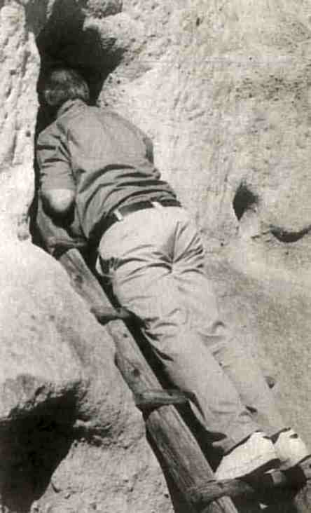 Watson Fuller investigating a new concept in student accommodation at the Bandelier National Monument. (Photo courtesy of R. Knott.)
The conference emphasized the importance of neutron techniques for the analysis of biological structure. Single crystal techniques will undoubtedly make a greater impact in the future. There is optimism in the reactor community that the future will be steady-state, and there could be a quantum leap in the use of pulsed neutron sources. The social acceptability of pulse neutron source technology will encourage greater innovation in equipment development.
The conference Organizing Committee was chaired by B. Schoenborn from the Life Sciences Division, Los Alamos National Lab and supported by grants from LANL, the IUCr, and the US DOE. All papers presented at the Conference will be published in a volume to be released in 1995.
Robert Knott