
Meeting report
Protein Society Meeting
The program of the annual meeting of the Protein Society featured advances in traditional and new techniques to characterize protein composition, sequence, conformation, dynamics, function and activity. Protein folding, systematic conformational analysis and reports of new structure determinations were emphasized. The meeting featured a superb and extensive exhibition of the latest in instrumentation for protein analysis. Techniques covered include X-ray, NMR, mass spectroscopy, electron diffraction, scanning tunneling microscopy, circular dichroism and fluorescence.
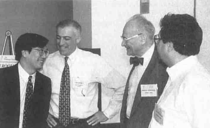 Peter Kim (MIT), Protein Society President, Joe Villafranca (Bristol Meyer, Squibb), Hal Scheraga (Cornell U.), Stein Moore Award winner for 1995, and Eugene Shakhnovich (Harvard U.) gather for the symposium session in honor of Scheraga. (Photo by WLD.)
At the July 1995 meeting, the Stein Moore award was presented to H. Scheraga in recognition of his pioneering work in predicting three-dimensional structure from protein sequence. Speakers in the symposium in Scheraga's honor included P. Kim and K. Wuthrich. (Scheraga reminded the audience that in 1949 when he began his work nobody knew what a protein looked like.)
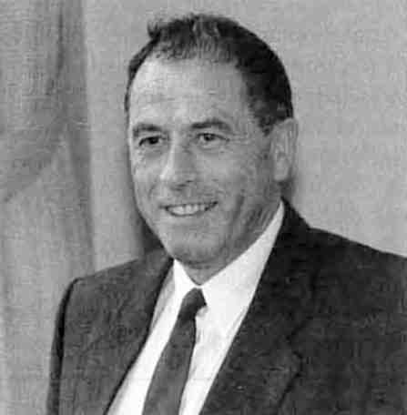 Kurt Wuthrich, pioneer in the field of high resolution macromolecular NMR studies, expounded on the complementary strength of NMR and X-ray technology at the 1995 Protein Society meeting. (Photo by WLD.)
Kim discussed 2, 3, and 4 stranded coiled coils for which T. Albers has done the X-ray structures. Wuthrich said that X-ray and NMR studies are complementary but also said crystal structures are not quite up to NMR standards. His paradigm structure has been Bovine pancreatic trypsine inhibitor (BPTI), an 88 amino acid peptide the X-ray structure of which was reported by Deisenhofer and Stiegleman in 1975. In 1992 Wuthrich obtained what he considers his current best set of NMR data on it. He reports that the side chains of the surface of the BPTI are different than they are observed in the crystal. In view of the fact that the NMR results indicate flexibility in the side chains of surface residues, it is remarkable that none of the positions correspond to the X-ray observations. Wuthrich concludes that this is due to an induced fit of the protein into the crystal lattice, despite few protein protein contacts in the BPTI crystals. All of the surface waters on BPTI are located with the NMR data and there is considerable correspondence between the water molecules that seem most ordered and well behaved in the X-ray determination and some of the highly detailed water structure established by the NMR study. The inter-conversion of disulfide bonds detected in the structure has a parallel in peptide crystal structures in which such a disorder has also been observed. Crystallographic analysis in which distinctions between static and dynamic disorder have been successfully delineated are rare. In hirudin not only surface residues but entire peptide strands exhibit multiple low energy conformations. Such information from NMR could augment X-ray structures in which poorly resolved or apparently entirely disordered terminal strands or internal loops are found. The NMR could identify a few most stable conformers that could be used to restrain a refinement of X-ray data effectively. Wuthrich also discussed time shared hydrogen bonds, water mediating protein DNA interactions and dynamic processes as monitored by NMR.
G. Petsko gave a brilliant talk including an introduction to Laue time resolved diffraction theory and application. A fourth dimension in the title of his talk referred to time. He used a multiple exposure photograph of Jack Nicklaus teeing off to illustrate protein dynamics. Greg reported that it has been possible to record 25,000 diffraction spots on elastase to 2 Å in 10 msec. Greg cited a Science article (June 1995) by Koshland and Stoddard on the citrate reaction that he considers the best dynamic study performed to date. Petsko applied the technique most recently to cytochrome P450 camphor (a different crystal form than the one well studied by T. Poulos). Greg showed four possible models for the intermediate and he showed that the final density was only compatible with one of the four.
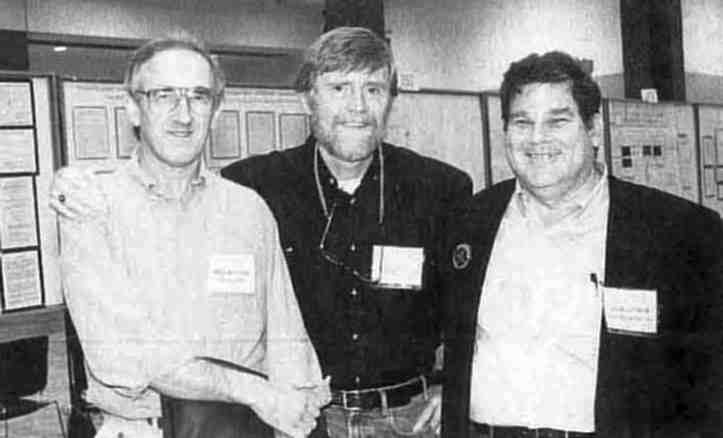 The Protein Society President elect, Brian Matthews (U. of Oregon), the ubiquitous editor, and smiling Ed Lattman (Johns Hopkins) compare notes on science, societies, technique and career choices. (Photo by WLD.)
D. Davies and collaborators crystallized a new HIV protein. The protein solubility needed for crystallization was only achieved after extensive mutational introduction of lysines and alanines to replace hydrophobic residues in the structure. The lysines were used to replace isolated hydrophobic residues and polyalanines for stretches of hydrophobic residues. The protein has a new fold characteristic of a growing family of endonucleases. P. Sigler was coauthor of a presentation about groEl and groES and the role of heat shock proteins or chaperonins in protein folding. Hsp60 plays a role in folding half a dozen different proteins. The chaperon appears to bea grooming device. GroEL and groES cooperate in the process of altering the protein fold of transient guests in the cavity of groEL.
C. Dobson gave a beautifully organized presentation of the use of data from many sources to probe the mechanism of protein folding. He compared his folding funnel cartoon with a representation of an energy profile having a band corresponding to the molten globule. Chris used lysozyme too illustrate the concept.
Other crystallographers on the very limited oral program, including W. Hol, D. Eisenberg, and J. Smith, presented eloquent expositions on complex protein systems. X-ray crystal structure determinations both old and new played a major role in a third of the 617 posters presented at the meeting. These included 25 reports of new protein crystal structures and a handful of others that described preparation of crystals suitable for X-ray analysis. At least 170 other posters included illustrations of three-dimensional structures (usually in color) taken from previous publications of X-ray crystal structure determinations. The X-ray structures were critical to the analysis of protein folding, mutational analysis,homology modeling, dynamic calculation, and/or mechanism of action that were the subjects of the research being reported. In some cases (20%) the original X-ray study is so well known that the authors saw no need to reference the source of the illustration. This was even the case in some posters when the authors were careful to provide extensive references to their own previous studies of the protein under analysis. Some of the authors who neglected to cite original crystallographic publications of the protein were in the vanguard that righteously insist that protein crystallographers be required to deposit their coordinates in the Brookhaven Database. There were about a dozen papers in which an ensemble of protein structures from the Brookhaven Database were the basis for development of new generalizations concerning the detail of protein structure. There were also a dozen reports of new protein structure determinations by NMR in solution and half a dozen new ab initio models proposed for protein folding.
The Protein Society Meeting continues to be the primary meeting bringing together all aspects of protein study. Protein crystallographers planning to attend the IUCr Congress in Seattle August 8-18, 1996 may want to extend their stay on the west coast of the US to include attendance at the Protein Society Meeting in San Diego August 3-7, 1996.
William L. Duax