
Meeting report
Let's Get Biophysical
The Thirty-Ninth Annual Meeting of the Biophysical Society, held in San Francisco in February 1995, offered a wealth of information on structure-function relationships and the application of X-ray crystal structure analysis to better understand these relationships. It was gratifying to learn from this meeting the tremendous impact that crystal structure analysis has upon biophysics.
Among the highlights of the meeting were many symposia devoted to new crystal structures. L. M. Gierasch and co-workers from J. Deisenhofer's lab in Dallas, Texas, reported on the 2.8 Å crystal structure of groES, a co-chaperone protein and proposed a mechanism by which groES and its co-chaperone protein, groEL, assist in protein folding. The Deisenhofer group also presented the crystal structure of the E. coli photolyase, a flavoprotein that recognizes and uses light to repair thymine dimers. This unique enzyme provides a model to better understand how DNA binding, electron transfer, and energy transduction are inter-related.
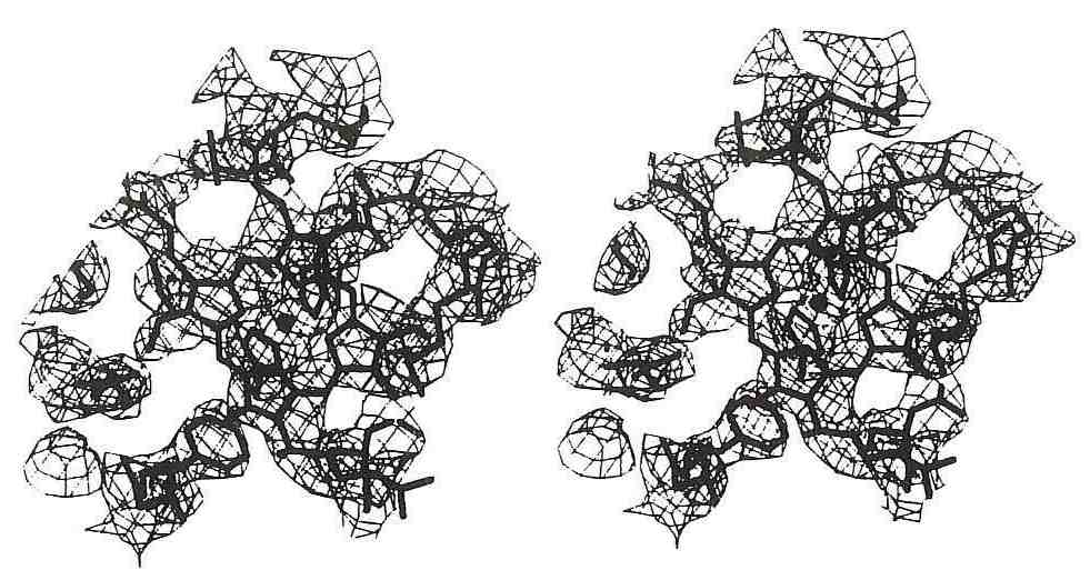 The crystal structures of cytochrome c oxidase from Bovine Heart (Tsukihara et al., Science 269, p. 1069) and P. denitrificans (Iwata et al., Nature 376, p. 660) are the largest membrane proteins yet crystallized at atomic resolution. The surroundings of the heme group in the enzyme are shown here. S. Yoshikawa, co-author of the Tsukihara paper, and H. Michel, co-author of the Iwata paper, will each address the XVII Congress in Seattle.
The transduction of light energy into chemical energy is an important and recurring theme in biophysics. Knowledge of the X-ray structures of the photosynthetic reaction centers, the first integral membrane protein crystal structures solved, has served to promote awareness of how light energy drives electron transfer and how this electron transfer is coupled to the formation of a transmembrane proton gradient. In 1988, H. Michel from the Max Planck Institute for Biophysics in Frankfurt, Germany and two of his collaborators, J. Deisenhofer and R. Huber, received the Nobel Prize in chemistry for the elucidation of the X-ray crystal structure of the photosynthetic reaction center from viridis.
Dr Michel presented his lab's recent work on the X-ray crystal structure of the photosynthetic reaction center from Rb. sphaeroides. The structure of this reaction center had been previously reported in an orthorhombic crystal form at a resolution of 2.8 Å by J. Allen and G. Feher (University of California, San Diego), and A. Chirino, H. Komiya, D. Rees, and T. Yeates (University of California, Los Angeles and CalTech). Michel reported an improved crystal structure for the Rb. sphaeroides reaction center based on a trigonal unit cell at a higher resolution (2.65 Å) and noted that the resolution in the trigonal form was enhanced because its lower packing density led to a finer sampling of the Fourier transform. The most striking difference in the trigonal structure is the inclusion of bound waters and a shift in the positioning of a bound quinone co-factor. Michel identified a chain of water molecules leading from the surface of the Reaction Center (RC) to the bound quinone acceptor, that may be involved in proton transfer. Results from other investigators using site-directed mutagenesis suggest that specific amino acid sidechains are important for the proton transfer activities of the RC. The results of site-directed mutagenesis investigations of the Rb. sphaeroides reaction centers were presented by H. Axelrod, M. Paddock, and S. Rongey (University of California, San Diego), and R. Isaacson described ENDOR studies of reacton center crystals. The importance of an internal water chain in the crystal structure of cytochrome f to proton translocation (W. Cramer, Purdue) and the role of water molecules in the methyl group transfer reactions performed by thymidylate synthase (R. Stroud, University of California, San Francisco) were described.
 Herbert Axelrod.
Other exciting presentations concerned membrane protein structure determinations. G. Scherther of the MRC in London captivated the audience with 7 Å three-dimensional images of the visual receptor, bovine rhodopsin obtained by cryoelectron microscopy. Progress on the crystallization and structure determination of two mitochondrial electron transfer chain proteins, cytochrome c reductase, 3.5 Å, and cytochrome c oxidase, 6.0 Å (S. H. Kim et al, Lawrence Berkeley Lab) was reported in the Poster Session. Structural reports on viral proteins included those of filamentous Pf1 virus, (3 Å fiber diffraction data, D. Liu, NY Public Health Research Institute and Yale University), and infectious adenovirus type 5 (J. Rux and R. Burnett, Wistar Institute).
In a symposium on proteins involved in contraction, I. Rayment described the structure of the nucleotide binding site in myosin subfragment-1 and R. Fletterick (University of California, San Francisco) spoke on kinesin motor domains. A poster session featured a report on the co-crystallization of a smooth muscle regulatory complex, tropomyosin-caldesmon at 17 Å resolution (E. Hnath, Rice University), a protocol to trap myosin subfragment 1 in a crosslinked form that can be crystallized (L. Riccelli, Washington State University), comparisons of calcium-free and calcium-bound forms of the N-terminal regulatory domain of troponin C (N. Strynadka, MRC, Edmonton) and structural changes that occur within actin upon binding profilin (K. J. Chik, Princeton University).
Reports of crystallographic studies of certain membrane active toxins included those on α-bungarotoxin bound to the nicotinic acetylcholine receptor using low angle X-ray scattering (H. Young, University of Connecticut), colicin E1 (P. Elkins, Purdue University), colicin 1A (M. Wiener, University of California, San Francisco), and diptheria toxin when bound to its endogenous inhibitor, ApUp (M. Weiss, University of California, Los Angeles). W. Meador (Baylor College of Medicine) described the structure of a calmodulin-like chimaeric protein bound to calmodulin-dependent protein kinase II-a, and R. Li (Johns Hopkins) spoke on the structure of the rat liver NADPH-quinone oxidoreductase, which protects cells from quinone carcinogens.
Structure-based investigations of proteins which bind metals included the study of uptake of iron into erythrocytes. This mechanism for iron uptake was deduced from the high resolution X-ray crystal structures of amphibian red cell L-ferritin (J. Trikha, University of Minnesota). Structure studies of heme proteins and porphyrins included chemically crosslinked hemoglobins (M. Berry, Rice University), an oxygen/carbon dioxide exchange protein from the hemolymph of the earthworm (K. Strand, University of Massachusetts) in which phases derived from 3-D cryo-electron microscopy were improved by multiple cycles of 6-fold averaging followed by phase extension, an examination of changes that occur upon photolysis of the bond between the heme iron and the carbon monoxide ligand in carbonmonoxymyoglobin (J. Berendzen, Los Alamos National Lab; I. Schlichting, Max Planck Institute for Medical Sciences; and G. Phillips, Rice University) and cooperative oxygen binding in blood clam hemoglobin in the liganded and unliganded form (W. Royer, University of Massachusetts).
Investigations of RNA and DNA structure included methods to predict the propensity of oligonucleotides to crystallize as A, B, or Z DNA (G. Schroth, Oregon State University, Corvall), a new class of A DNA crystal structures, hexanucleotides which pack in a C2221 space group (B. Mooers, Oregon State University), and a detailed comparison of the structures of a B-DNA oligonucleotide implicated in sequence-specific DNA bending (M. Young, Wesleyan University). One highlight of a symposium on catalytic RNA was H. Pley's (Stanford University) talk on the 3-D X-ray crystal structure determination of a hammerhead RNA-DNA ribozyme inhibitor complex.
Structural aspects of protein-nucleic acid interactions also included the structures of DNA polymerase processivity factors (J. Kuriyan, Rockefeller University), an extension of the resolution of the catabolite gene activator protein-DNA complex (G. Parkinson. Rutgers University), and the localization of the binding site of the carboxy-terminal domain of RNA polymerase II determined from e1ectron crystallography (G. Meredith, Stanford University).
Herbert L. Axelrod